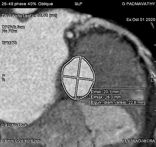
Before you undergo TAVR, your cardiologist will recommend various diagnostic tests to assess your overall health and risk factors. One of the most common tests that doctors prescribe is a CT angiogram.
Before you undergo TAVR, your cardiologist will recommend various diagnostic tests to assess your overall health and risk factors. One of the most common tests that doctors prescribe is a CT angiogram.
TAVR is a new procedure that has been gaining popularity in recent years. It’s used for the treatment of aortic stenosis. It involves the use of a catheter to replace a damaged aortic valve with a biological tissue valve.
Aortic valve stenosis is a serious condition that doesn’t just affect the aortic valve. It reduces or blocks the blood supply to the aorta. That, in turn, restricts blood flow to vital organs, including the brain, kidneys, and liver.
If left untreated, aortic stenosis can lead to severe complications, including:
All these conditions can threaten a patient’s well-being and, ultimately, result in death.
Also, it’s worth noting that aortic stenosis can worsen over time, progressing from stage A (at risk) to stage D (symptomatic severe). As the disease progresses, the prognosis becomes worse, too.
As the above graph shows, the likelihood of a patient’s survival rapidly decreases after the onset of aortic stenosis symptoms. Despite advancements in medical science, the outlook for patients with severe aortic stenosis remains poor.
That makes it crucial for aortic stenosis patients to seek treatment at the earliest. Timely diagnosis and treatment can help prevent unfavorable outcomes like arrhythmias and strokes. Also, early intervention increases the likelihood of success of treatments like aortic valve replacement and aortic valve repair.
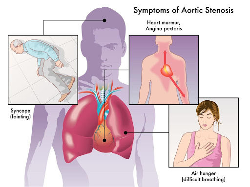
If you’re at risk of aortic stenosis due to congenital heart defects or a genetic predisposition, watch out for symptoms like heart murmur, chest pain, and shortness of breath. Also, make sure you regularly visit a cardiologist to get your heart’s chambers and valves evaluated.
Severe aortic stenosis is characterized by significant damage to the aortic valve. Whether asymptomatic or symptomatic, the condition poses the risk of heart failure, blood clots, and even death. That makes it crucial for patients to receive treatment for aortic valve stenosis.
Patients with severe aortic stenosis need surgery to repair or replace the aortic valve. Typically, your cardiologist will recommend one of the following procedures:
It’s a minimally invasive procedure that involves inserting a catheter with a balloon tip into an artery in the arm or groin. Once the catheter reaches the aortic valve, the balloon is inflated to expand the narrowed valve opening. Thereafter, the balloon is deflated and removed, along with the catheter.
Balloon valvuloplasty is a suitable aortic stenosis treatment option for infants and children. In adults, the aortic valve can narrow again after the procedure. However, it can be used as temporary aortic valve stenosis treatment for adults waiting for valve replacement.
It’s a type of open heart surgery where a cardiac surgeon removes the damaged aortic valve and replaces it with a mechanical valve or biological tissue valve. Biological tissue valves are made from cow, pig, or human heart tissue. They usually disintegrate over time and may need to be surgically replaced.
While mechanical valves are more durable, they increase the risk of bleeding and thromboembolic complications. Patients with mechanical valves will need to take blood thinners to minimize the risk of blood clots.
In some cases, the patient’s pulmonary valve is used to replace the aortic valve, and a mechanical valve is secured in place of the pulmonary valve. Also known as Ross Procedure, the surgery is more complex and time-consuming.
![]()
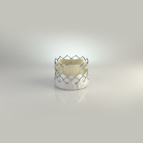
TAVR is a minimally invasive procedure suitable for patients who can’t undergo open-heart aortic valve replacement due to comorbidities or old age. In TAVR, the surgeon inserts a catheter through a tiny incision in the patient’s arm or groin. The catheter is used to transport and install a replacement valve made from cow or pig tissue to the aortic valve region. We’ll cover TAVR in more detail later in this article.
Mild or moderate aortic valve stenosis may not need to be treated with surgery right away. However, it’s worth noting that aortic stenosis will worsen over time, and the outlook is poor for patients in the severe stage.
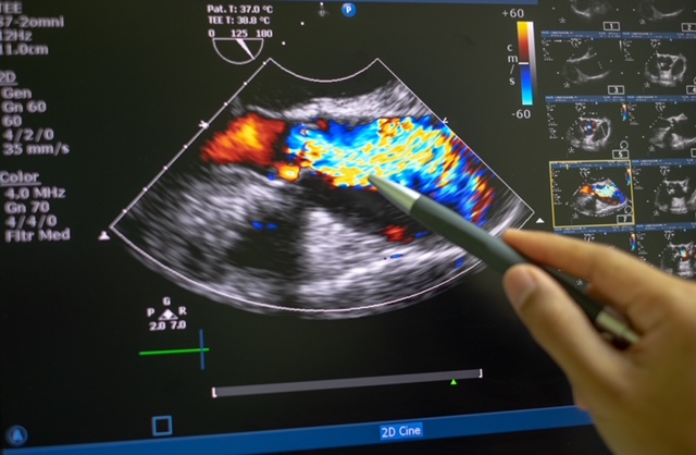
That makes it crucial for patients with moderate aortic stenosis to visit their doctor for routine evaluation and follow-ups. Your doctor might prescribe tests like echocardiogram and chest X-rays to assess the extent of valve damage.
Additionally, your doctor will recommend healthy lifestyle changes to slow down the progression of aortic stenosis. These include:
At this stage, your doctor will also focus on the management of underlying conditions, such as hypertension and chronic kidney disease, that increase the risk of aortic stenosis. They’ll prescribe suitable medications to treat these conditions.

Depending on your overall health and medical history, you can talk to your doctor and explore the possibility of undergoing aortic valve replacement (open-heart or transcatheter). Make sure you visit the doctor for regular check-ups to monitor the progression of aortic stenosis.
Medical treatment and lifestyle changes can help manage the symptoms and slow down the progression of aortic valve stenosis. However, surgery is the only way to improve a patient’s longevity and quality of life.
If you’re looking for aortic stenosis treatment, you can choose from the following surgeries:
Balloon valvuloplasty involves inserting a catheter with a balloon tip into an artery and inflating the balloon tip to widen the aortic valve opening. The procedure is minimally invasive and better suited for children and infants. It can also be used as a temporary fix for patients with severe aortic stenosis awaiting valve replacement.
Aortic valve replacement involves replacing the damaged aortic valve with a mechanical or biological tissue valve during open-heart surgery. If a patient has several comorbidities, they may not be an ideal candidate for AVR. In such cases, they can opt for a less invasive procedure called transcatheter aortic valve replacement.
Just like balloon valvuloplasty, TAVR also involves inserting a catheter with a balloon tip into a blood vessel. The catheter is guided to the aortic valve region and used to transport the replacement valve.
The type of surgery you’ll need depends on your current health, medical history, and the severity of aortic stenosis. It’s wiser to talk to your doctor to explore your options and choose the right surgery.
Transcatheter aortic valve replacement (TAVR) is a minimally invasive procedure used for the treatment of aortic stenosis. It’s an alternative to traditional aortic valve replacement, which requires open-heart surgery.
The minimally invasive nature of TAVR makes it suitable for aortic stenosis patients who can’t undergo open-heart surgery due to comorbidities, such as lung and kidney diseases. Also known as transcatheter aortic valve implantation (TAVI), the procedure can relieve symptoms like chest pain and shortness of breath and improve a patient’s quality of life.
Unlike open-heart surgery, TAVR doesn’t require the surgeon to make a long incision on the patient’s chest. Instead, they insert a thin tube (catheter) into the patient’s artery through a tiny incision in the arm or groin area. The catheter is fitted with a balloon tip and guided to the aortic valve region.
Once the catheter reaches the aortic valve, a replacement valve (made from cow or pig tissue) is passed through it. The balloon tip inflates to securely place the replacement valve in the correct position (in place of the aortic valve). After the successful implantation of the replacement valve, the catheter and balloon tip are withdrawn.
You might receive sedatives before the procedure to help you relax. Also, you’ll likely have to spend the night in the ICU after undergoing TAVR. It helps your treatment team monitor your vitals and manage any complications that might arise after the procedure.
While TAVR is a fairly safe procedure, it comes with the following risks:
Most patients have to take anticoagulant medications to prevent the formation of blood clots. Also, your doctor might prescribe antibiotics to protect the replacement valve from infection. It’s wiser to talk to your doctor and assess the benefits and risks of TAVR before undergoing the procedure.

Aortic valve stenosis is a serious disease that can lead to heart failure, strokes, and even death if left untreated. That makes timely diagnosis and treatment of the condition crucial. However, treatment of aortic stenosis depends on its severity.
Typically, the following parameters are used to determine the severity of aortic stenosis:
Depending on the pressure gradient and blood flow parameters, aortic stenosis is graded as follows:
NF-HG is the most prevalent type of aortic stenosis and has well-established management protocols. Patients with NF-HG are also ideal candidates for aortic valve replacement. While LF-LG is fairly rare, it’s often associated with a poor prognosis.
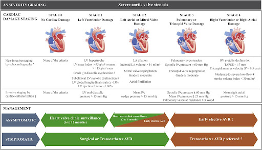
Additionally, depending on progression, heart valve disease can be categorized into the following four stages:
Doctors use a variety of diagnostic tests to evaluate the aforementioned parameters and determine the severity of aortic stenosis. If you experience symptoms like chest pain, heart murmur, or palpitation, it’s crucial to reach out to an experienced cardiologist and get the right treatment for aortic valve stenosis.
Early diagnosis of aortic valve stenosis is crucial to prevent severe complications, such as arrhythmias, heart failure, stroke, and death. Also, it can help administer timely treatment, thus improving the patient’s prognosis and quality of life.
That’s why cardiologists use a series of tests to diagnose aortic valve stenosis and its underlying cause. When you visit the doctor, they’ll start by asking you about your symptoms and medical history. Also, they ask whether your family has a history of cardiovascular ailments. Next, they’ll use a stethoscope to detect the presence of the characteristic aortic stenosis murmur.
Additionally, your doctor will use one or more of the following tests for the complete diagnosis:
Additionally, your doctor might recommend tests like cardiac catheterization and chest X-ray to get a complete picture of your cardiac health and plan the right course of treatment.

The heart is a vital organ of the human body. It pumps blood throughout your body and keeps you alive. It comprises cardiac muscle tissue. That means the heart can contract and relax to squeeze blood out of the heart and into your body. It supplies blood to all organs, including the brain, kidneys, liver, and more.
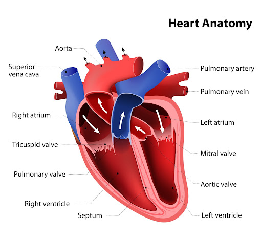
The heart has four chambers divided by two walls called septa. The upper chambers are called the atria, and the lower chambers are called the ventricles.
The atria receive blood from the veins and pump it into the ventricles through openings called valves. The tricuspid valve separates the right atrium from the right ventricle, and the mitral valve separates the left atrium from the left ventricle. There’s also a pulmonary valve that sits between the right ventricle and the pulmonary artery.
Then there’s the aortic valve located between the left ventricle and the aorta. It prevents blood from leaking back into the left ventricle during the contraction phase of the heart’s pumping cycle.
When your heart beats, it squeezes blood out through an opening in each chamber called an aortic valve into either a large artery (aorta) or one of its branches, the carotid arteries, before heading back down to smaller arteries throughout your body. When the heart pumps blood to other organs, oxygen will be picked up by red blood cells as they pass through capillaries.
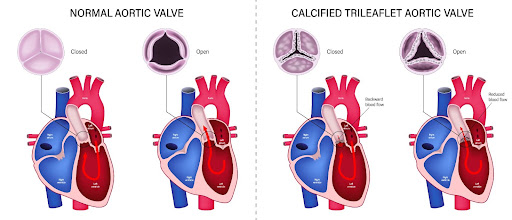
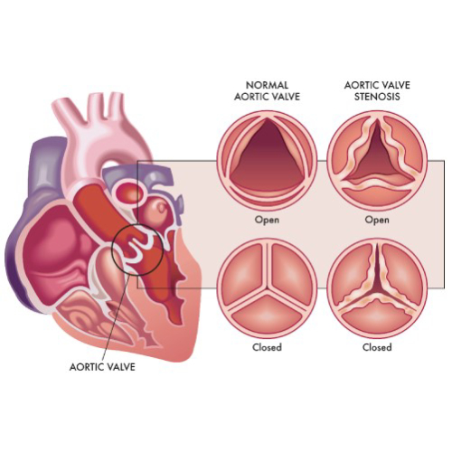
The aortic valve is one of the four valves in the heart. It is a flap of tissue that keeps blood flowing in one direction. It is located in the middle of the heart, between the left ventricle and the aorta, the main artery that supplies blood throughout the body. The aortic valve is a semilunar valve, meaning the flaps of the valve are crescent-shaped.
The aortic valve comprises three sections called leaflets or cusps. Each cusp is made of collagen. Some people are born with only two cusps in the aortic valve. In such cases, it is known as a bicuspid aortic valve.
The aortic valve opens when the heart contracts or squeezes and closes when the heart relaxes. It lets oxygen-rich blood flow from the left ventricle into the aorta for circulation throughout your body.
The aortic valve is crucial because it ensures that blood keeps flowing in the right direction from the heart. It prevents the backflow of blood from the aorta into your left ventricle.
If the aortic valve doesn’t work properly, it can cause serious problems. A leaky or stiffened valve won’t open and close properly, which means that some blood flows backward into the left ventricle. It puts extra pressure on the heart and can lead to heart failure or stroke if severe enough.
The aortic valve is a crucial part of the heart that keeps blood flowing in the right direction throughout the body. The two primary diseases that affect the aortic valve are:
In aortic valve stenosis the cusps (or flaps) of the aortic valve become stiff and thick, thus narrowing the valve opening. That, in turn, restricts blood flow from the heart’s left ventricle to the aorta and the rest of the body.
Aortic valve stenosis is usually caused by pre-existing conditions, such as congenital heart defects, aortic valve calcification, and rheumatic fever. Chronic ailments, such as hypertension, endocarditis, and diabetes, and treatments like radiation therapy to the chest can also increase the risk of aortic valve stenosis.
In aortic valve regurgitation, the flaps don’t close properly, causing a backflow of blood into the left ventricle. As with aortic valve stenosis, this condition is also caused by pre-existing ailments that affect the heart.
Common symptoms of aortic valve disease include heart murmur, chest pain, dizziness, fatigue, irregular heartbeat, and shortness of breath. Treatment for these conditions depends on the severity of the disease and its underlying cause. If left untreated, both aortic valve stenosis and aortic valve regurgitation can lead to heart failure and other complications.
| According to the CDC, nearly 2.5% of the US population suffers from heart valve diseases. These conditions are more common in older people, with 13% of people born before 1943 experiencing them. Also, 1% to 2% of the US population is affected by the bicuspid aortic valve, with the condition being more common in men. |
If you experience any of the aforementioned symptoms, reach out to an experienced cardiologist for proper diagnosis and treatment.
Aortic stenosis is a heart valve disease affecting the aortic valve that connects the heart’s left ventricle (lower chamber) to the aorta (main artery). The aortic valve is responsible for ensuring that blood flows in one direction throughout the body.
In aortic valve stenosis, the aortic valve flaps (or cusps) become stiff or thick due to calcium buildup in the valve or other underlying causes. It causes the valve opening to become narrow and restricts blood flow to the aorta. That, in turn, reduces or blocks the blood supply to vital organs, such as the liver, kidney, brain, etc.
Symptoms of aortic valve stenosis include:
Depending on the underlying cause, there are two types of aortic stenosis – congenital and acquired. Congenital aortic stenosis is present from birth due to a defect in the valve’s formation. Acquired stenosis develops after birth due to other heart-related ailments, such as hypertension, endocarditis, rheumatic fever, and aortic valve calcification.
The risk of aortic stenosis increases in older people. Also, people who have received radiation therapy to the chest are more vulnerable to the condition.
Treatment for aortic valve stenosis depends on the underlying cause and the severity of the condition. Many patients need surgery to repair the valve. If left untreated, aortic stenosis can lead to heart failure, stroke, arrhythmias, and even death. Therefore, it’s crucial to diagnose aortic valve stenosis and treat the condition at the earliest.
Copyright © 2023, Dr. Raghu. All rights reserved.
+91 95424 75650

+91 95424 75650