Heart Valve disease / Dr Raghu

Aortic-stenosis.jpg

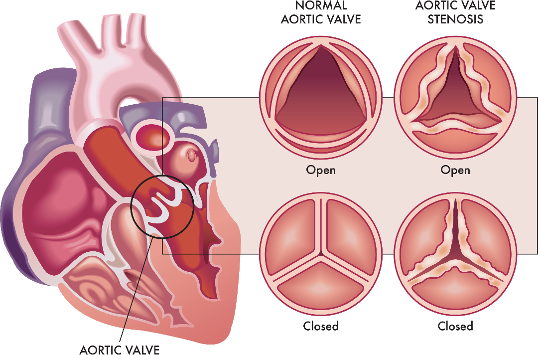
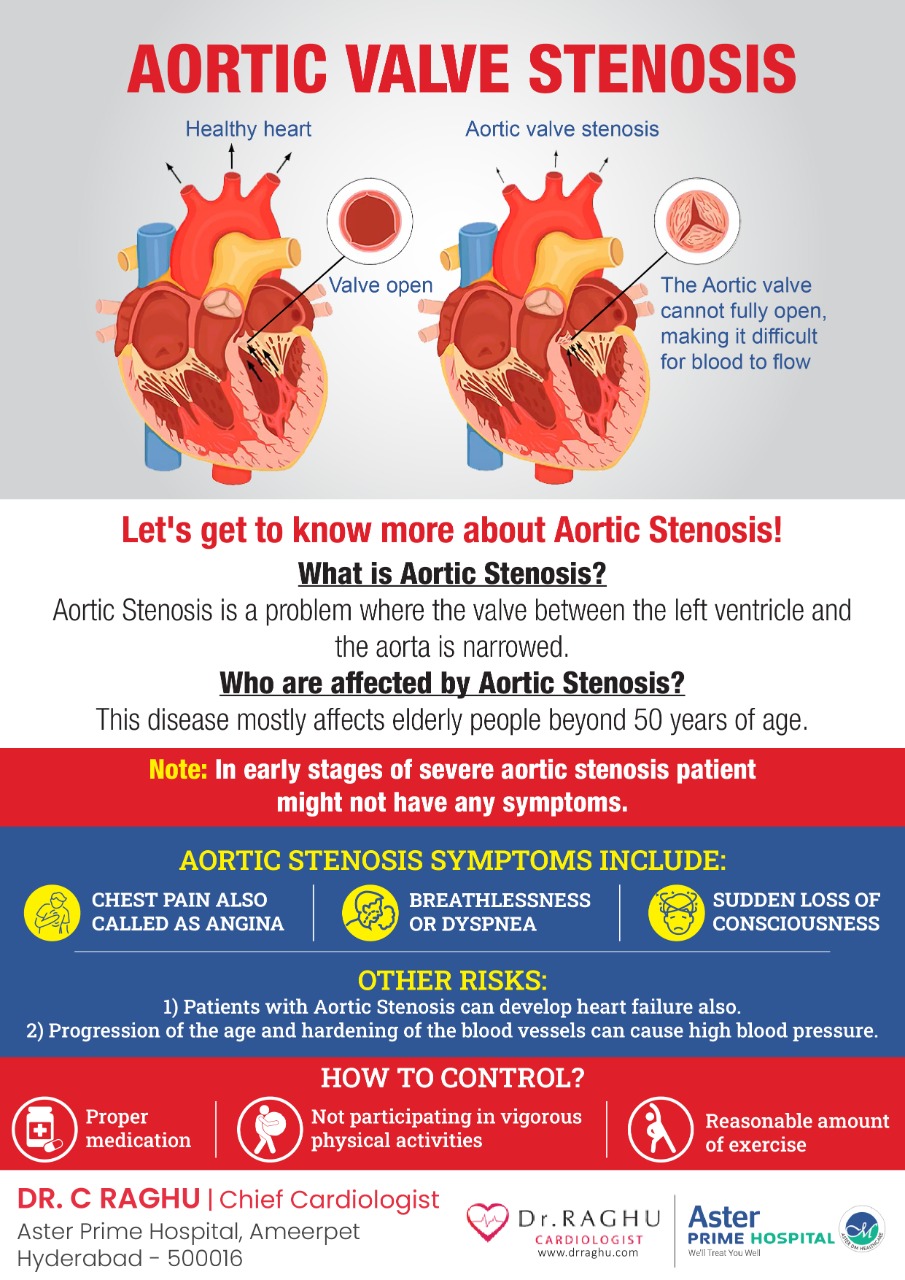
What is Aortic stenosis?

Aortic stenosis is a disease where the valve between left ventricle and aorta is narrowed. Normally the left ventricle is the chamber which pumps blood to the entire body through the aortic valve. So if the aortic valve is narrowed either due to infection or age related degeneration it is called aortic stenosis. This is a disease of the elderly people usually beyond 50 years of age.

What are the symptoms of aortic stenosis?

When the disease is severity is mild people usually do not experience any symptoms. But is the disease is severe – even though the disease is severe in early phases the patient might not have symptoms. 

So, early stages of aortic stenosis patient might not have any symptoms. Where as in the advanced stage of severe aortic stenosis the patients can develop can develop chest pain also called angina in medical terminology. So chest pain which increases on walking or any other form of exertion and gets relieved on rest or stopping of that activity is called angina. So patients with aortic stenosis because is unable to pump as per the requirements of the body (due to aortic valve narrowing) they experience angina. 

The other symptom is breathlessness – on walking or on lying down flat the patient develops difficulty in breathing. This is referred to as dyspnea in medical terminology. So when ever a patient is having a fixed blood supply to various organs without increasing as per their needs there is pooling of blood in the heart. This pooled blood in the heart “reverses back” into the lungs which is responsible of breathlessness.

Finally in advanced stages of aortic stenosis patients develop a sudden loss of consciousness with spontaneous recovery. These episodes of loss of consciousness are also called as syncope in medical terminology.

So the predominant symptoms of aortic stenosis are chest pain, breathlessness and sudden loss of consciousness.

At the same time patients with aortic stenosis will develop an impaired function of the heart also called heart failure. This heart failure need to necessarily present in severe heart failure but can also be seen in intermediate or moderately severe aortic stenosis. 

Aortic stenosis and high blood pressure 

Patients with aortic stenosis have reduced supply of blood to various organs of the body. Because of this it was believed that patients with aortic stenosis tend to have low blood pressure. This is not true regarding the current epidemic of aortic stenosis we are currently seeing. Currently most of the aortic stenosis patients are elderly in their 60s, 70s and 80s of age. So these patients because of the progression of the age and hardening of the blood vessels they develop high blood pressure or Hypertension in medical terminology. So patients with aortic stenosis are not spared from high blood pressure contrary to what we were believing till date and what we are seeing is a scenario of aortic stenosis patients having high blood pressure levels.

What is the impact of this high blood pressure on a patient with severe aortic stenosis?

Patients with high blood pressure and severe aortic stenosis develop a faster progression of the disease severity. So a patient of aortic stenosis with uncontrolled blood pressure can have a severe aortic stenosis at a much earlier age. 

How can patients with aortic stenosis control their blood pressure?

People with aortic stenosis and high blood pressure need to control their blood pressure using 3-4 different types of medicines. A good control of blood pressure is one of the first steps in retarding the progression of aortic stenosis. 

Can people with aortic stenosis do exercise? 

People with aortic stenosis tend to have a fixed cardiac output. This means – the aortic valve is narrowed and this narrowing limits the blood supply to various organs of the body. When there is a reduction in blood supply to various organs of the body – the first to be affected is the brain. This causes syncope or sudden loss of consciousness. 

Exercise AS

So when a person with severe aortic stenosis exercises vigorously then there is a reduction of blood supply to the brain causing sudden unconsciousness. This problem happens in people with an advanced or severe aortic stenosis. So people with severe or advanced aortic stenosis are advised not to participate in vigorous physical activity such as running, jogging or weight lifting etc. 

But at the same time as we all know for the control of BP, blood sugar and cholesterol and effective control of heart failure are important steps for retarding the progression of aortic stenosis. So a mild to moderate severe intensity exercise is advised for control of the various co-morbidities in aortic stenosis. But at the same time a vigorous or severe intensity exercise is definitely not to be performed. Severe intensity exercise or competitive sports is a contra indication for aortic stenosis patients in medical terminology.

Aortic stenosis

Aortic Stenosis Symptoms (Telugu)

Book Online Consultaion


    Aortic Stenosis : Symptoms Blog

    DOWNLOAD PDF

    Subscribe the Hearty Life Blogs


      disease-condtions-mitral_stenosis.jpg

      Mitral stenosis is a form of valvular heart disease caused by the narrowing of the mitral valve. Mitral valve lies between the left atrium and left ventricle of the heart which is made up of two flaps of tissue called leaflets. It opens when the blood flows from left atrium and left ventricle and closes immediately to prevent the backward flow of the blood. The defective valve fails to either open or close completely.

      The most common cause of mitral stenosis is an infection called rheumatic fever. It is an inflammatory condition that usually starts with strep throat and leads to permanent damage of heart valves. Rheumatic fever is now rare in developed countries, but the prevalence is still high in developing countries. It may scar the mitral valve and if left untreated, mitral stenosis may lead to severe heart complications. Mitral stenosis can be classified into three types – mild, moderate and severe depending on the severity.

      Symptoms:

      The progression of is slow, and the symptoms generally appear after 20 to 40 years after an episode of rheumatic fever. However, an individual with mitral stenosis may feel fine or have minimal symptoms for decades. They include:

      • Shortness of breath, especially during physical effort or when you lie down
      • Chest discomfort or chest pain
      • Fatigue and weakness, especially during increased physical activity and during pregnancy
      • Swollen feet or legs
      • Heart palpitations – sensations of a rapid, fluttering heartbeat
      • Dizziness or fainting
      • Coughing up blood
      • Thromboembolic complications such as stroke

      Mitral stenosis symptoms may worsen due to any activity that can cause an increase in the heart rate. The pressure which is built up in the heart due to mitral stenosis causes fluid buildup in the lungs. The symptoms of mitral stenosis usually appear between ages of 15 to 40 years. But they can appear in any age or even during childhood.

      The signs that can be found during general examination include:

      • Heart murmur observed using stethoscope during clinical examination
      • Fluid buildup in the lungs
      • Irregular heart rhythms (arrhythmias)

      Causes:

      • Rheumatic fever: The major cause of mitral stenosis is rheumatic fever. Rheumatic fever is a complication of strep throat which can damage mitral valve by thickening or fusing the valves.
      • Other causes include:
      • Calcium deposits: People of older age can develop calcium deposits. This leads to calcification of the mitral valve leaflets resulting in mitral valve stenosis.
      • Congenital heart disease: Some babies may be born with a narrowed mitral valve, that may lead to mitral stenosis.

      Risk factors:

      The individuals with the following conditions are at risk of mitral stenosis:

      • Infective endocarditis
      • Endomyocardial fibroelastosis
      • Malignant carcinoid syndrome
      • Systemic lupus erythematosus
      • Whipple disease
      • Rheumatoid arthritis

      Diagnosis:

      The diagnosis of mitral stenosis could follow an invasive or non-invasive method.

      The noninvasive procedures include:

      Electrocardiogram (ECG):  In this procedure, the electrodes are attached to pads on patients’ skin to measure electrical impulses from the heart which provides information about heart rhythm. The patient is either made to walk on a treadmill or pedal a stationary bike during an Electrocardiogram (ECG) to see how the heart responds to exertion.

      Echocardiogram: The echocardiogram is a very useful tool to assess the mitral stenosis etiology, morphology, severity, and treatment intervention.

      Two types of echocardiogram are performed which include:

      • Transthoracic echocardiogram: This test is used to confirm the diagnosis of mitral stenosis. In this procedure, the sound waves are directed to patients’ heart from a transducer held near the chest which produces video images of heart in motion.
      • Transesophageal echocardiogram: In this procedure a small transducer is attached to the end of a tube which is inserted into esophagus. This provides a closer look at the mitral valve when compared to regular echocardiogram.
      • Chest X-ray: The chest X-ray is used to observe the size of the heart size, prominent main pulmonary arteries, dilatation of the upper pulmonary veins, and displacement of the esophagus by an enlarged left atrium. If the condition is severe there could be enlargement of all the chambers, pulmonary arteries, and pulmonary veins. The chest X-ray also helps to identify the condition of lungs.

      The invasive procedures include:

      Cardiac catheterization: Cardiac catheterization is an invasive procedure and is performed when the noninvasive tests are inconclusive or when there is a no correlation between noninvasive tests and clinical findings. It involves threading a thin tube (catheter) through a blood vessel in the patients arm or groin to the coronary artery in the heart and injecting dye through the catheter to make the artery visible on an X-ray. This provides a detailed picture of your heart.

      These cardiac tests help in distinguishing mitral valve stenosis from other heart conditions, including other mitral valve conditions. These tests also help reveal the cause of your mitral valve stenosis and whether the valve can be repaired.

      Prevention:

      The patients with mild mitral stenosis without any symptoms generally do not require an immediate treatment, but are monitored continuously. Medications are prescribed to ease the workload of the heart and to regulate its rhythm, thus reducing the symptoms. The following medications may be prescribed:

      • Diuretics to reduce fluid accumulation in the body or lungs
      • Blood thinners (anticoagulants) such as daily intake of aspirin to help prevent blood clots
      • Beta blockers or calcium channel blockers to slow your heart rate and allow your heart to fill more effectively
      • Anti-arrhythmics to treat atrial fibrillation or other rhythm disturbances
      • Antibiotics to prevent a recurrence of rheumatic fever if it is an underlying cause for the condition

      The Treatment Involves Surgical or Nonsurgical Procedures

      Nonsurgical procedures:

      Percutaneous balloon mitral valvuloplasty

      In this procedure, which is also called balloon mitral valvotomy, a soft, thin tube (catheter) tipped with a balloon is inserted in an artery of the patients arm or groin and guided into the narrowed valve. Once in position, the balloon is inflated to widen the valve, improving the blood flow. The balloon is then deflated, and the catheter with balloon is removed.

      Surgical options include:

      • Commissurotomy: An open-heart surgery is performed to remove calcium deposits and other scar tissues to clear the valve passageway.
      • Mitral valve replacement: Mitral valve replacement is considered if it cannot be repaired. In mitral valve replacement, the damaged valve is removed and is replaced with a mechanical valve or a biological tissue valve.

      Prevention:

      The prevention of mitral stenosis is largely dependent on preventing the occurrence of rheumatic fever, which is the major cause of the condition. However, if one acquires rheumatic fever, the following measures can help live a healthy life.

      • Limit intake of salt.
      • Maintain a healthy body weight.
      • Decrease caffeine intake.
      • Seek prompt medical attention.
      • Cut back on alcohol.
      • Perform regular exercise.
      • Go for regular checkups.

      MITRAL VALVE DISEASE – Mitral Stenosis( in Telugu)

      Book Online Consultaion


        Mitral Stenosis Blog

        DOWNLOAD PDF

        Subscribe the Hearty Life Blogs


          +91 95424 75650

          Call us now if you are in a medical emergency need, we will reply swiftly and provide you with a medical aid.


          +91 95424 75650


          Call us now if you are in a medical emergency need, we will reply swiftly and provide you with a medical aid.


          Dr. Raghu | Heart Specialist in Hyderabad
          Yashoda Hospitals, Sardar Patel Rd, behind Hari Hara Kala Bhavan, Kummari Guda, Shivaji Nagar, Secunderabad, Telangana 500003


          Copyright © 2023, Dr. Raghu. All rights reserved.

          [gtranslate]