Resources / Dr Raghu

aortic-stenosis-1.jpg

Why Should One Undergo Early Treatment for Aortic Stenosis?

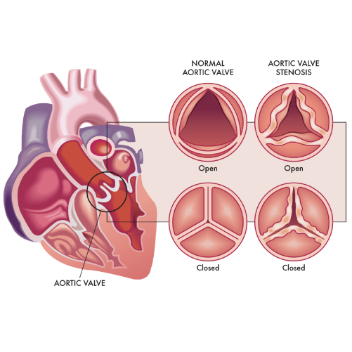
Aortic valve stenosis is a serious condition that doesn’t just affect the aortic valve. It reduces or blocks the blood supply to the aorta. That, in turn, restricts blood flow to vital organs, including the brain, kidneys, and liver.

If left untreated, aortic stenosis can lead to severe complications, including:

  • Heart failure
  • Blood clots and stroke
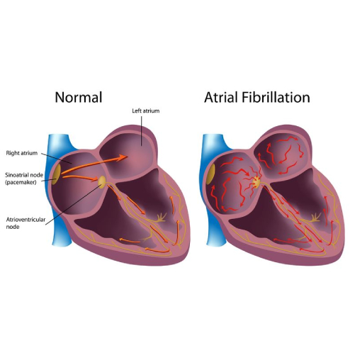
  • Arrhythmias (irregular heartbeat)
  • Bleeding
  • Infections of the heart (including endocarditis)

All these conditions can threaten a patient’s well-being and, ultimately, result in death.

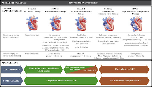
Also, it’s worth noting that aortic stenosis can worsen over time, progressing from stage A (at risk) to stage D (symptomatic severe). As the disease progresses, the prognosis becomes worse, too.

As the above graph shows, the likelihood of a patient’s survival rapidly decreases after the onset of aortic stenosis symptoms. Despite advancements in medical science, the outlook for patients with severe aortic stenosis remains poor.

That makes it crucial for aortic stenosis patients to seek treatment at the earliest. Timely diagnosis and treatment can help prevent unfavorable outcomes like arrhythmias and strokes. Also, early intervention increases the likelihood of success of treatments like aortic valve replacement and aortic valve repair.

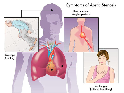
If you’re at risk of aortic stenosis due to congenital heart defects or a genetic predisposition, watch out for symptoms like heart murmur, chest pain, and shortness of breath. Also, make sure you regularly visit a cardiologist to get your heart’s chambers and valves evaluated.

What Is the Treatment for Severe Aortic Stenosis?

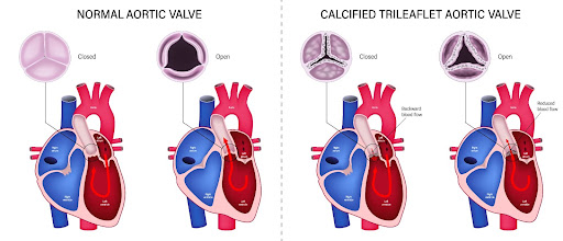
Severe aortic stenosis is characterized by significant damage to the aortic valve. Whether asymptomatic or symptomatic, the condition poses the risk of heart failure, blood clots, and even death. That makes it crucial for patients to receive treatment for aortic valve stenosis.

Patients with severe aortic stenosis need surgery to repair or replace the aortic valve. Typically, your cardiologist will recommend one of the following procedures:

Balloon Valvuloplasty

It’s a minimally invasive procedure that involves inserting a catheter with a balloon tip into an artery in the arm or groin. Once the catheter reaches the aortic valve, the balloon is inflated to expand the narrowed valve opening. Thereafter, the balloon is deflated and removed, along with the catheter.

Balloon valvuloplasty is a suitable aortic stenosis treatment option for infants and children. In adults, the aortic valve can narrow again after the procedure. However, it can be used as temporary aortic valve stenosis treatment for adults waiting for valve replacement.

Aortic Valve Replacement

It’s a type of open heart surgery where a cardiac surgeon removes the damaged aortic valve and replaces it with a mechanical valve or biological tissue valve. Biological tissue valves are made from cow, pig, or human heart tissue. They usually disintegrate over time and may need to be surgically replaced.

While mechanical valves are more durable, they increase the risk of bleeding and thromboembolic complications. Patients with mechanical valves will need to take blood thinners to minimize the risk of blood clots.

In some cases, the patient’s pulmonary valve is used to replace the aortic valve, and a mechanical valve is secured in place of the pulmonary valve. Also known as Ross Procedure, the surgery is more complex and time-consuming.

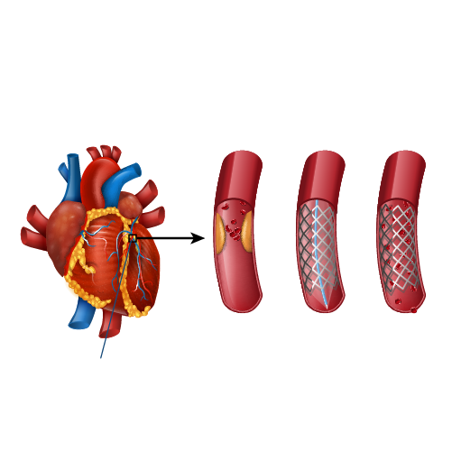
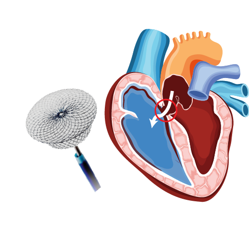
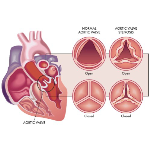
Transcatheter Aortic Valve Replacement (TAVR)

TAVR is a minimally invasive procedure suitable for patients who can’t undergo open-heart aortic valve replacement due to comorbidities or old age. In TAVR, the surgeon inserts a catheter through a tiny incision in the patient’s arm or groin. The catheter is used to transport and install a replacement valve made from cow or pig tissue to the aortic valve region. We’ll cover TAVR in more detail later in this article.

What Is Medical Treatment for Moderate Aortic Stenosis?

Mild or moderate aortic valve stenosis may not need to be treated with surgery right away. However, it’s worth noting that aortic stenosis will worsen over time, and the outlook is poor for patients in the severe stage.

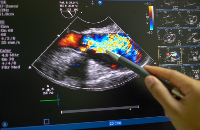
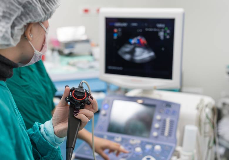
That makes it crucial for patients with moderate aortic stenosis to visit their doctor for routine evaluation and follow-ups. Your doctor might prescribe tests like echocardiogram and chest X-rays to assess the extent of valve damage.

Additionally, your doctor will recommend healthy lifestyle changes to slow down the progression of aortic stenosis. These include:

  • Avoiding tobacco smoking and alcohol consumption
  • Getting enough sleep
  • Following a daily exercise routine
  • Eating a balanced, low-fat diet

At this stage, your doctor will also focus on the management of underlying conditions, such as hypertension and chronic kidney disease, that increase the risk of aortic stenosis. They’ll prescribe suitable medications to treat these conditions.

Depending on your overall health and medical history, you can talk to your doctor and explore the possibility of undergoing aortic valve replacement (open-heart or transcatheter). Make sure you visit the doctor for regular check-ups to monitor the progression of aortic stenosis.

Surgery for Aortic Stenosis 

Medical treatment and lifestyle changes can help manage the symptoms and slow down the progression of aortic valve stenosis. However, surgery is the only way to improve a patient’s longevity and quality of life.

If you’re looking for aortic stenosis treatment, you can choose from the following surgeries:

  • Balloon valvuloplasty
  • Aortic valve replacement (AVR)
  • Transcatheter aortic valve replacement (TAVR)

Balloon valvuloplasty involves inserting a catheter with a balloon tip into an artery and inflating the balloon tip to widen the aortic valve opening. The procedure is minimally invasive and better suited for children and infants. It can also be used as a temporary fix for patients with severe aortic stenosis awaiting valve replacement.

Aortic valve replacement involves replacing the damaged aortic valve with a mechanical or biological tissue valve during open-heart surgery. If a patient has several comorbidities, they may not be an ideal candidate for AVR. In such cases, they can opt for a less invasive procedure called transcatheter aortic valve replacement.

Just like balloon valvuloplasty, TAVR also involves inserting a catheter with a balloon tip into a blood vessel. The catheter is guided to the aortic valve region and used to transport the replacement valve. 

The type of surgery you’ll need depends on your current health, medical history, and the severity of aortic stenosis. It’s wiser to talk to your doctor to explore your options and choose the right surgery.

TAVR for Aortic Stenosis

Transcatheter aortic valve replacement (TAVR) is a minimally invasive procedure used for the treatment of aortic stenosis. It’s an alternative to traditional aortic valve replacement, which requires open-heart surgery.

The minimally invasive nature of TAVR makes it suitable for aortic stenosis patients who can’t undergo open-heart surgery due to comorbidities, such as lung and kidney diseases. Also known as transcatheter aortic valve implantation (TAVI), the procedure can relieve symptoms like chest pain and shortness of breath and improve a patient’s quality of life.

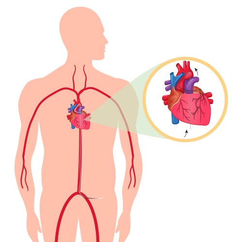
Unlike open-heart surgery, TAVR doesn’t require the surgeon to make a long incision on the patient’s chest. Instead, they insert a thin tube (catheter) into the patient’s artery through a tiny incision in the arm or groin area. The catheter is fitted with a balloon tip and guided to the aortic valve region.

Once the catheter reaches the aortic valve, a replacement valve (made from cow or pig tissue) is passed through it. The balloon tip inflates to securely place the replacement valve in the correct position (in place of the aortic valve). After the successful implantation of the replacement valve, the catheter and balloon tip are withdrawn.

You might receive sedatives before the procedure to help you relax. Also, you’ll likely have to spend the night in the ICU after undergoing TAVR. It helps your treatment team monitor your vitals and manage any complications that might arise after the procedure.

While TAVR is a fairly safe procedure, it comes with the following risks:

  • Heart attack
  • Stroke
  • Bleeding
  • Infection
  • Arrhythmias

Most patients have to take anticoagulant medications to prevent the formation of blood clots. Also, your doctor might prescribe antibiotics to protect the replacement valve from infection. It’s wiser to talk to your doctor and assess the benefits and risks of TAVR before undergoing the procedure.

Book Online Consultaion


    Treatment of Aortic Stenosis – Blog

    DOWNLOAD PDF

    Subscribe the Hearty Life Blogs


      Echocardiogram-standard.jpeg

      Grading Severity of Aortic Stenosis

      Aortic valve stenosis is a serious disease that can lead to heart failure, strokes, and even death if left untreated. That makes timely diagnosis and treatment of the condition crucial. However, treatment of aortic stenosis depends on its severity.

      Typically, the following parameters are used to determine the severity of aortic stenosis:

      • Pressure gradient – High gradient (HG; >/=40 mm Hg) or low gradient (LG; <40 mm Hg)
      • Blood flow – Normal flow (NF; SVi>35 ml/m2) or low flow (LF; SVi<35ml/m2)
      • Left ventricular ejection fraction (LVEF) – Preserved (>/=50%) or reduced (<50%)

      Depending on the pressure gradient and blood flow parameters, aortic stenosis is graded as follows:

      • Normal flow-low gradient (NF-LG)
      • Normal flow-high gradient (NF-HG)
      • Low flow-high gradient (LF-HG)
      • Low flow-low gradient (LF-LG)

      NF-HG is the most prevalent type of aortic stenosis and has well-established management protocols. Patients with NF-HG are also ideal candidates for aortic valve replacement. While LF-LG is fairly rare, it’s often associated with a poor prognosis.

      Aortic-valve-stenosis 

      Additionally, depending on progression, heart valve disease can be categorized into the following four stages:

      • Stage A (At risk) – Characterised by the presence of risk factors
      • Stage B (Progressive) – Mild or moderate valve disease with no noticeable symptoms
      • Stage C (Asymptomatic severe) – Severe valve damage with no noticeable symptoms
      • Stage D (Symptomatic severe) – Severe valve disease with noticeable symptoms

      Doctors use a variety of diagnostic tests to evaluate the aforementioned parameters and determine the severity of aortic stenosis. If you experience symptoms like chest pain, heart murmur, or palpitation, it’s crucial to reach out to an experienced cardiologist and get the right treatment for aortic valve stenosis.

      How Does One Diagnose Aortic Stenosis – ECG, ECHO, TEE, or CT aortogram?

      Early diagnosis of aortic valve stenosis is crucial to prevent severe complications, such as arrhythmias, heart failure, stroke, and death. Also, it can help administer timely treatment, thus improving the patient’s prognosis and quality of life.

      That’s why cardiologists use a series of tests to diagnose aortic valve stenosis and its underlying cause. When you visit the doctor, they’ll start by asking you about your symptoms and medical history. Also, they ask whether your family has a history of cardiovascular ailments. Next, they’ll use a stethoscope to detect the presence of the characteristic aortic stenosis murmur.

      Additionally, your doctor will use one or more of the following tests for the complete diagnosis:

      • ECG (Electrocardiogram) – It’s one of the most preliminary tests that evaluate the heart’s electrical activity and helps doctors identify an irregular heartbeat and other abnormalities.
      • Echo (Echocardiogram) – It uses sound waves to generate images of the beating heart. It helps doctors examine how blood flows through each valve and determine the severity of aortic stenosis.
      • TEE (Transesophageal echocardiogram) – It’s a special type of echocardiogram in which an ultrasound probe is inserted into the esophagus and directed closer to the heart. It helps doctors take a closer look at the aortic valve and identify the underlying cause of aortic stenosis.
      • CT aortogram – It’s used to evaluate the blood supply to the upper body and identify conditions such as atherosclerosis.

      Echocardiography (ultrasound) machine. doppler of aortic stenosis

      Additionally, your doctor might recommend tests like cardiac catheterization and chest X-ray to get a complete picture of your cardiac health and plan the right course of treatment.

      Book Online Consultaion


        Diagnosing Aortic Stenosis – Blog

        DOWNLOAD PDF

        Subscribe the Hearty Life Blogs


          Aotic-Valve.jpg

          Basics of the Heart

          The heart is a vital organ of the human body. It pumps blood throughout your body and keeps you alive. It comprises cardiac muscle tissue. That means the heart can contract and relax  to squeeze blood out of the heart and into your body. It supplies blood to all organs, including the brain, kidneys, liver, and more. 

          The heart has four chambers divided by two walls called septa. The upper chambers are called the atria, and the lower chambers are called the ventricles.

          The atria receive blood from the veins and pump it into the ventricles through openings called valves. The tricuspid valve separates the right atrium from the right ventricle, and the mitral valve separates the left atrium from the left ventricle. There’s also a pulmonary valve that sits between the right ventricle and the pulmonary artery.

          Then there’s the aortic valve located between the left ventricle and the aorta. It prevents blood from leaking back into the left ventricle during the contraction phase of the heart’s pumping cycle.

          When your heart beats, it squeezes blood out through an opening in each chamber called an aortic valve into either a large artery (aorta) or one of its branches, the carotid arteries, before heading back down to smaller arteries throughout your body. When the heart pumps blood to other organs, oxygen will be picked up by red blood cells as they pass through capillaries.

          What Is the Aortic Valve, and Why Is It Important?

          Aotic Valve

          The aortic valve is one of the four valves in the heart. It is a flap of tissue that keeps blood flowing in one direction. It is located in the middle of the heart, between the left ventricle and the aorta, the main artery that supplies blood throughout the body. The aortic valve is a semilunar valve, meaning the flaps of the valve are crescent-shaped.

          The aortic valve comprises three sections called leaflets or cusps. Each cusp is made of collagen. Some people are born with only two cusps in the aortic valve. In such cases, it is known as a bicuspid aortic valve.

          The aortic valve opens when the heart contracts or squeezes and closes when the heart relaxes. It lets oxygen-rich blood flow from the left ventricle into the aorta for circulation throughout your body. 

          The aortic valve is crucial because it ensures that blood keeps flowing in the right direction from the heart. It prevents the backflow of blood from the aorta into your left ventricle.

          If the aortic valve doesn’t work properly, it can cause serious problems. A leaky or stiffened valve won’t open and close properly, which means that some blood flows backward into the left ventricle. It puts extra pressure on the heart and can lead to heart failure or stroke if severe enough.

          Which Diseases Affect the Aortic Valve?

          The aortic valve is a crucial part of the heart that keeps blood flowing in the right direction throughout the body. The two primary diseases that affect the aortic valve are:

          • Aortic valve stenosis
          • Aortic valve regurgitation

          In aortic valve stenosis the cusps (or flaps) of the aortic valve become stiff and thick, thus narrowing the valve opening. That, in turn, restricts blood flow from the heart’s left ventricle to the aorta and the rest of the body.

          Aortic valve stenosis is usually caused by pre-existing conditions, such as congenital heart defects, aortic valve calcification, and rheumatic fever. Chronic ailments, such as hypertension, endocarditis, and diabetes, and treatments like radiation therapy to the chest can also increase the risk of aortic valve stenosis.

          In aortic valve regurgitation, the flaps don’t close properly, causing a backflow of blood into the left ventricle. As with aortic valve stenosis, this condition is also caused by pre-existing ailments that affect the heart.

          Common symptoms of aortic valve disease include heart murmur, chest pain, dizziness, fatigue, irregular heartbeat, and shortness of breath. Treatment for these conditions depends on the severity of the disease and its underlying cause. If left untreated, both aortic valve stenosis and aortic valve regurgitation can lead to heart failure and other complications.

          According to the CDC, nearly 2.5% of the US population suffers from heart valve diseases. These conditions are more common in older people, with 13% of people born before 1943 experiencing them. Also, 1% to 2% of the US population is affected by the bicuspid aortic valve, with the condition being more common in men.

          If you experience any of the aforementioned symptoms, reach out to an experienced cardiologist for proper diagnosis and treatment.

          Aortic Stenosis: What Is It?

          symptoms of Aortic Stenosis

          Aortic stenosis is a heart valve disease affecting the aortic valve that connects the heart’s left ventricle (lower chamber) to the aorta (main artery). The aortic valve is responsible for ensuring that blood flows in one direction throughout the body.

          In aortic valve stenosis, the aortic valve flaps (or cusps) become stiff or thick due to calcium buildup in the valve or other underlying causes. It causes the valve opening to become narrow and restricts blood flow to the aorta. That, in turn, reduces or blocks the blood supply to vital organs, such as the liver, kidney, brain, etc.

          Symptoms of aortic valve stenosis include:

          • A whooshing or swishing heart sound (known as aortic stenosis murmur)
          • Chest pain or tightness
          • Fatigue and dizziness (particularly after physical activity)
          • Shortness of breath
          • Palpitations

          Depending on the underlying cause, there are two types of aortic stenosis – congenital and acquired. Congenital aortic stenosis is present from birth due to a defect in the valve’s formation. Acquired stenosis develops after birth due to other heart-related ailments, such as hypertension, endocarditis, rheumatic fever, and aortic valve calcification.

          The risk of aortic stenosis increases in older people. Also, people who have received radiation therapy to the chest are more vulnerable to the condition. 

          Treatment for aortic valve stenosis depends on the underlying cause and the severity of the condition. Many patients need surgery to repair the valve. If left untreated, aortic stenosis can lead to heart failure, stroke, arrhythmias, and even death. Therefore, it’s crucial to diagnose aortic valve stenosis and treat the condition at the earliest.

          Book Online Consultaion


            Understanding Aortic Stenosis – Blog

            DOWNLOAD PDF

            Subscribe the Hearty Life Blogs


              artifiacial-sweetener.jpg

              If you’re trying to lose weight or control your blood glucose levels, you’re likely looking for ways to cut your intake of refined sugar. Artificial sweeteners, such as saccharin, sucralose, and xylitol, are a popular replacement for refined sugar.

              heart disease

              However, there’s always been controversy around the potential harm these sweeteners can cause in the long run. For instance, long-term consumption can make an individual more susceptible to type 2 diabetes and weight gain. That, in turn, can increase the risk of cardiovascular diseases.

              A recent study by the French National Institute of Health and Medical Research substantiates that claim. The report, published in The BMJ, found a higher rate of cardiovascular disease events in participants who consumed higher levels of artificial sweeteners.

              A Closer Look at the Study

              The study involving more than 170,000 French citizens commenced in 2009 with participants aged 18 years and older. The average age of the participants was 42 years, and a majority of them were females.

              The researchers regularly collected information about the participants’ dietary habits, including their consumption of artificial sweeteners. Nearly 37% of participants used such sweeteners and consumed 42.46 mg/day on average.

              The artificial sweeteners consumed by the participants included the following:

              • Aspartame
              • Acesulfame potassium
              • Sucralose
              • Saccharin
              • Steviol glycosides (and more)

              At the end of the study, the researchers uncovered a direct correlation between the intake of artificial sweeteners and the risk of heart-related ailments. Higher consumers of such sweeteners had a 9% higher incidence of cardiovascular diseases and an 18% higher incidence of cerebrovascular diseases.

              While aspartame was associated with an increased risk of strokes, acesulfame potassium and sucralose were linked with a higher risk of coronary heart disease.

              What Does That Mean For You?

              The recent study of French nationals echoes previous claims about the harmful effects of artificial sweeteners. Long-term consumption of these sweeteners can be detrimental to your cardiac health.

              However, that doesn’t mean you should completely steer clear of them. Occasional intake within the permissible limits won’t pose any threat to you. The recommended limit depends on your body weight and the type of sweeteners.

              If you’re looking to lose weight, get your blood sugar under control, or embrace healthy eating habits, your best bet is to consult a certified nutritionist. Also, it’s a good idea to consider natural alternatives to refined sugar. These include allulose and stevia. 

              Additionally, consider staying away from processed and packaged foods. Many of these items contain artificial sweeteners. Also, they don’t have a high nutritional value and could increase your cholesterol levels.

              In Conclusion

              Regular consumption of artificial sweeteners like aspartame and sucralose can have an adverse effect on your heart. They increase your risk of coronary heart disease, strokes, and other ailments. 

              If you’re looking for ways to minimize your refined sugar intake, consider natural alternatives like stevia. Also, consult a doctor or nutritionist to understand the side effects of different refined sugar replacements.

              Dr. C Raghu is an eminent cardiologist with decades of experience. He’s helped countless patients lead healthier lives. If you or anyone you know has been diagnosed with a cardiovascular ailment, feel free to reach out to Dr. Raghu today.

              Book Online Consultaion


                Artificial Sweeteners and the Risk of Heart Disease – Blog

                DOWNLOAD PDF

                Subscribe the Hearty Life Blogs


                  Aspirin.jpg

                  Doctors devise a customized treatment plan when a patient is diagnosed with a cardiac ailment. Typically, treatment for heart diseases includes a combination of medicines, lifestyle changes, and in some cases, surgery.

                  One of the most commonly prescribed medicines for heart-related ailments is aspirin. In this article, we’ll delve deeper into the use of aspirin for heart disease and analyze its pros and cons. Let’s get started.

                  What Is Aspirin?

                  Aspirin is one of the most widely used over-the-counter drugs in the world. It comes in several forms, but the most common way to take it is as a pill. It belongs to a specific class of medications called nonsteroidal anti-inflammatory drugs (NSAIDs).

                  Aspirin

                  Besides being an effective anti-inflammatory medication and pain reliever, aspirin has anticoagulant properties. That means it can help prevent heart disease and stroke when taken regularly over time. 

                  Aspirin works by preventing blood clots from forming in blood vessels through its effects on platelets, which are cells that help blood clot. There is some controversy about whether or not you should take aspirin to prevent heart disease, given its potential side effects (such as stomach ulcers and bleeding). 

                  But if your physician recommends it as part of your overall treatment plan, it may be worth considering.

                  How Aspirin Helps Prevent Heart Attacks, Strokes, and Death

                  Aspirin’s ability to reduce the risk of heart attacks, strokes, and cardiovascular death stems from its effects on several pathways in the body. The drug helps to prevent blood clots by inhibiting an enzyme called cyclooxygenase (COX). 

                  COX is responsible for producing prostaglandins, which cause inflammation and pain. By reducing the production of these substances, aspirin also lowers blood pressure and reduces inflammation in the body.

                  Additionally, aspirin is an antiplatelet drug that helps prevent blood clotting by preventing platelets from sticking together. That means taking aspirin regularly can help keep your heart healthy by reducing the chances of a stroke or heart attack caused by a blood clot forming inside your arteries or veins.

                  Aspirin Use for Primary Prevention

                  Aspirin can be used to minimize the risk of cardiovascular disease in at-risk patients. This is known as primary prevention. According to the American College of Cardiology and the American Heart Institute, a low dose (75 to 100 mg/day) of aspirin can help with the primary prevention of atherosclerotic cardiovascular diseases among at-risk adults in the age group of 40 to 70 years. However, if you have a bleeding disorder, aspirin may not be the best fit for you.

                  Aspirin Use for Secondary Prevention

                  Besides protecting at-risk patients, aspirin can also be given to patients who’ve suffered a heart attack or stroke, received a coronary angioplasty, or undergone a coronary bypass. The anticoagulant properties of aspirin help minimize the risk of cardiovascular events in the future. This is known as secondary prevention.

                  Risks Associated With Taking Aspirin

                  The most common side effects of aspirin are related to the stomach and intestines. Prolonged use can lead to gastrointestinal bleeding and stomach ulcers. That, in turn, can cause abdominal cramping, pain, and indigestion.

                  Should You Take Aspirin to Prevent Heart Disease?

                  Your doctor is in the best position to decide whether you should take aspirin for your cardiac health. You’ll benefit from taking aspirin if you have a history of heart attacks or strokes. However, you should consult your doctor for the proper dosage and duration of treatment.

                  Also, if you are pregnant or breastfeeding, talk to your doctor. Aspirin is classified as Pregnancy Category B by the Food and Drug Administration (FDA). That means it is safe for use during pregnancy in animal studies and causes in humans. However, data are limited, so consult with your doctor before taking aspirin while pregnant.

                  It’s worth noting that aspirin is a blood thinner and can cause bleeding and bruising. You shouldn’t take it with other blood thinners, such as warfarin (Coumadin), heparin, clopidogrel (Plavix), theophylline (Theo-Dur), or dipyridamole (Persantine).

                  Aspirin you should avoid aspirin if you have the following:

                  • A bleeding disorder or are taking other medications that increase your risk of bleeding
                  • Ulcers on your stomach or duodenum

                  In Conclusion

                  If you have been diagnosed with heart failure or other cardiac ailments, your doctor may recommend aspirin therapy to help prevent heart attacks and strokes. It’s an anticoagulant that prevents the formation of blood clots. Make sure you talk to your doctor about the potential risks and side effects.

                  Dr. C Raghu is an accomplished cardiologist who’s been practicing for decades. If you have concerns about taking aspirin for heart disease, feel free to consult Dr. Raghu today.

                  Book Online Consultaion


                    Aspirin for the Prevention of Heart Disease – Blog

                    DOWNLOAD PDF

                    Subscribe the Hearty Life Blogs


                      Care-After-Coronary-Angiography.jpg

                      Many people have heart disease, but not all of them know it. Doctors use a variety of tests to identify cardiac ailments and their underlying causes. Coronary angiography is one of the most common diagnostic tests that doctors use.

                      While it’s considered a safe and minimally invasive procedure, coronary angiography comes with a few risks. You can read more about the potential risks and complications in our previous blog.

                      The good news is that you can avoid side effects with proper care after the procedure. If you’ve recently had coronary angiography or are going through one soon, here is what you need to do after the procedure:

                      Avoid Heavy Physical Activity

                      It’s important to rest after coronary angiography. Avoid lifting heavy objects or resuming your exercise routine right after the procedure. Make sure you get plenty of sleep and let your body and mind relax for at least a week after angiography. Ask your doctor if you’re unsure when to hit the gym or start exercising.

                      Embrace Healthy Habits

                      Avoid cigarettes for at least a few days if you are a smoker. Smoking can cause spasms in the coronary arteries, leading to a heart attack or stroke after your angiography. Also, avoid alcohol consumption, and make sure you eat a balanced diet.

                      If you are a coffee drinker or have high blood pressure, avoiding all caffeine products (coffee, tea) and salt for several days following your procedure may be a good idea. The most important thing is to listen carefully to what your doctor tells you about caring after coronary angiography to avoid any other complications during recovery or later down the road.

                      Be Careful About Removing the Bandage

                      A crucial step to caring after coronary angiography is to avoid removing the bandage until the morning of the second day after the procedure. Also, you should not remove it yourself. Let someone else help you remove it. You must consult your doctor or nurse before removing the bandage.

                      Expect Soreness and Pain for a Few Days

                      Your arm or leg (where the catheter was inserted) might feel sore for a few days following a coronary angiogram. The discomfort you feel is usually caused by the catheter or dye used for the test. 

                      The catheter may have been left in place too long, leading to irritation and inflammation. Or it could be a reaction to the dye that was used to take images of your arteries during the procedure. It can also happen if you’re allergic to any component of either one.

                      In most cases, the pain and soreness should subside after a few days. If the pain persists or the arm/leg becomes numb, it’s a good idea to reach out to your doctor. Also, if you have a fever or trouble breathing, call your doctor right away. 

                      If you are bleeding from the incision site, apply direct pressure with a gauze dressing until the bleeding stops. Don’t remove the dressing unless instructed by medical staff at the hospital.

                      Conclusion

                      Coronary angiography is a safe and effective procedure that can give you a better understanding of your heart health. If you follow your doctor’s instructions, you’ll be on the road to recovery in no time.

                      Dr. C Raghu is a renowned cardiologist who has treated thousands of patients with cardiac ailments. If you have any concerns or questions about coronary angiography, feel free to reach out to Dr. Raghu today.

                      Book Online Consultaion


                        Care After Coronary Angiography – Blog

                        DOWNLOAD PDF

                        Subscribe the Hearty Life Blogs


                          patent-foramen.jpg

                          The heart is a vital organ that supplies blood to the muscles and tissues and keeps your body running. In our previous articles, we’ve discussed common conditions that affect the heart, such as congestive heart failure and atrial fibrillation. Take a look at our blog for more information.

                          While much is discussed about these common ailments, other conditions like patent foramen ovale (or PFO) can also affect the heart. So, what exactly is PFO? And how does it affect your cardiac health? In this article, we’ll explore the ins and outs of this lesser-known condition.

                          What is Patent Foramen Ovale?

                          Patent Foramen Ovale (PFO) is a hole in the heart that can be congenital or acquired. The foramen ovale is a flap-like opening between the heart’s upper chambers. It usually closes within three to six months after birth and becomes a part of your heart’s septum.


                          patent foramen ovale

                          But if that doesn’t happen, you may have a PFO. In most cases, a PFO is present at birth (congenital). However, one can also develop it as an adult due to injury, infection, or inflammation of the tissue around the heart (acquired). 

                          Should You Be Worried About Patent Foramen Ovale?

                          If you’ve been diagnosed with a patent foramen ovale, you’re likely wondering if it poses a threat to your cardiac health. Fortunately, this condition is usually asymptomatic and does not require treatment. However, it can be a risk factor for stroke and heart attack in some people.

                          PFO can also be the underlying cause of other conditions, such as:

                          • Hypoxemia (low blood oxygen)
                          • Pulmonary hypertension
                          • Paradoxical embolism
                          • Migraine
                          • Atrial Fibrillation

                          That makes it crucial to stay in touch with your doctor and monitor your heart health to keep these conditions at bay.

                          Signs and Symptoms of PFO

                          If you’re lucky, a PFO will cause no noticeable symptoms. However, if it results in a drop in your blood oxygen levels, you may experience a variety of symptoms, including:

                          • Headaches
                          • Dizziness
                          • Chest pain (angina)
                          • Shortness of breath 
                          • Fatigue or weakness that does not go away with rest
                          • Feeling faint

                          What Are the Treatment Options for Patent Foramen Ovale?

                          Treatment options for a PFO include:

                          • Device closure procedure, is necessary only for patients who have had a stroke or heart attack caused by the condition
                          • Anticoagulant or antiplatelet medication for those with a complex morphology PFO
                          • Watchful waiting if you have no symptoms (that means you will be monitored regularly for any problems caused by your condition)

                          If you experience symptoms, your doctor may recommend lifestyle changes or medication as treatment options.

                          In Conclusion

                          Now you know what a patent foramen ovale is and why it’s so important to get checked out if you have symptoms. If you are diagnosed with a PFO, your doctor may recommend treatment based on your symptoms and medical history. That said, PFO does not always require treatment. Some people have been living with it for years without any issues at all.

                          Dr. C Raghu is an eminent cardiologist who specializes in interventional cardiology. If you or anyone you know has been diagnosed with a patent foramen ovale, feel free to consult Dr. Raghu to explore your treatment options.

                          Book Online Consultaion


                            Patent Foramen Ovale – Blog

                            DOWNLOAD PDF

                            Subscribe the Hearty Life Blogs

                              dr. raghu

                              Dr. RAGHU

                              MD, DM, FESC, FACC, FSCAI

                              Cardiology Coronary, Vascular and

                              Structural Interventions


                              Conditions & Diseases

                              Coronary angioplasty

                              Angioplasty

                              Coronary angioplasty

                              Aortic Stenosis

                              Coronary angioplasty

                              Atrial Fibrillation

                              Coronary angioplasty

                              Atrial Septal Defect


                              View More Services


                              cardiac_catheterization1.jpg

                              In our previous blogs, we’ve discussed various tests doctors use to monitor cardiac health and diagnose conditions like coronary artery disease and heart failure.

                              It’s worth noting that commonly used diagnostic tests like coronary angiography rely on a procedure called cardiac catheterization. In this blog, we’ll discuss the procedures in greater detail to help you understand the benefits and risks.

                              Cardiac catheterization is a procedure in which doctors insert a thin, flexible tube called a catheter into your heart through an artery. 

                              What Is Cardiac Catheterization?

                              Cardiac catheterization is a procedure that uses a thin tube (called a catheter) to examine the heart and blood vessels. The catheter is inserted into a blood vessel in the groin or arm. Thereafter, it’s advanced through the blood vessels and into the heart to measure pressures within different chambers of your heart.

                              cardiac catheterization

                              The test lets doctors see if there’s any blockage in the blood vessels that supply blood to your heart. Also, they can assess whether the cardiac muscles and valves are in proper working condition. Also, it may be used to insert wires (called stents) in coronary arteries if they become narrow due to plaque buildup or after an injury.

                              To perform balloon angioplasty, the doctor inflates a tiny balloon at one end of the catheter inside one of the blocked coronary arteries. It creates more space for oxygen-rich blood flow through your coronary arteries and helps improve symptoms like chest pain caused by a heart attack

                              How Does Cardiac Catheterization Work?

                              In this procedure, a doctor inserts the catheter into a blood vessel in your arm or groin. The catheter is guided through the blood vessels to the heart, where it’s used to look for blockages or other problems in the coronary arteries that supply oxygen-rich blood to your heart muscle.

                              Afterward, a dye is injected into your bloodstream from an intravenous (IV) line connected to your vein. It helps doctors see how well blood flows through different parts of your heart on X-rays taken during the procedure.

                              Why Do Doctors Use Cardiac Catheterization?

                              Cardiac catheterization can be used to diagnose or treat various heart ailments. It can help your doctor determine if your heart is getting enough blood and if there are any blockages in the arteries that supply blood to the rest of your body. Also, it can help identify any problems with the heart muscles and valves.

                              What Are the Risks of Cardiac Catheterization?

                              While the risk of complications from cardiac catheterization is low, there are some things you should know before your procedure. For example, you might also experience bleeding or bruising around where the cut was made.

                              The most severe risk is having a stroke due to excess pressure in the brain caused by increased blood flow due to plaque shaving off from arteries. Other risks include allergic reactions to dyes, bleeding at the puncture site where the catheter was inserted, and infection.

                              Conclusion

                              Cardiac catheterization is a standard procedure that can be done in a cath lab or hospital. It allows doctors to check for problems with your heart and blood vessels. Ask your doctor any questions about how long it will take and whether there are any risks associated with cardiac catheterization before scheduling an appointment for this test.

                              Dr. C Raghu is an accomplished cardiologist with decades of experience in interventional cardiology. If you or anyone you know is experiencing symptoms of cardiac ailments, book an appointment with Dr. Raghu today.

                              Book Online Consultaion


                                Cardiac Catheterization – Blog

                                DOWNLOAD PDF

                                Subscribe the Hearty Life Blogs


                                  CT-Coronary-Angiography-1.jpg

                                  Doctors use various diagnostic tests to assess cardiac health and diagnose conditions like coronary artery disease and heart failure. While a coronary angiogram is commonly used to identify these conditions, your doctor can prescribe other tests like CT coronary angiography. 

                                  In our previous blogs, you can read more about the use cases and risks of coronary angiography. In this article, we’ll delve deeper into CT coronary angiography and understand why it’s crucial for people with cardiac ailments.

                                  What Is CT Coronary Angiography?

                                  CT coronary angiography is a type of CT scan that uses special dyes to evaluate the heart’s blood vessels. It can detect blockages in the coronary arteries that supply blood to the heart. It can identify blockages that are too small for other methods, such as a regular angiogram or an electrocardiogram (ECG), to pick up.

                                  A CT coronary angiography scan aims to find any abnormal areas in your coronary arteries that could be causing stenosis (narrowing) or occlusion (blockage). It helps doctors identify underlying heart-related ailments and determine the right course of treatment.

                                  How Does CT Coronary Angiography Work?

                                  The test is usually done in a hospital or clinic. To begin with, the doctor or radiologist will start an intravenous (IV) line to administer the contrast dye. As the contrast flows through your veins, it will appear on the scan as bright white areas on a black background. It’ll help the doctor see your heart and coronary arteries. 

                                  The test usually lasts about 30 minutes to one hour. The CT coronary angiogram procedure involves two scans: one with diastolic blood flow and another with systolic blood flow

                                  When Is CT Coronary Angiography Used?

                                  CT coronary angiography is used to diagnose a heart attack and evaluate blood flow in the heart. Also, doctors use it to detect blockages in arteries that supply blood to the heart muscle.

                                  What Are the Risks of CT Coronary Angiography?

                                  While CT coronary angiography is a non-invasive and safe procedure, it can cause the following side effects:

                                  • You may have an allergic reaction to the contrast dye.
                                  • Your body will be exposed to high levels of radiation, which can increase your risk of developing cancer.
                                  • You might have to pay out of pocket because the test isn’t always covered by insurance.
                                  • You might experience more anxiety and stress if the scans indicate blockages in your arteries.

                                  What Should I Expect When Having CT Coronary Angiography?

                                  If you are planning to get a CT coronary angiography, you should be prepared for the following:

                                  • The radiologist will ask you to change into a hospital gown.
                                  • The radiologist will give you an injection of a contrast dye.
                                  • You will lie on a table, and the table will move into the CT scanner.
                                  • The scan is painless and takes about 20 minutes.

                                  Conclusion

                                  CT coronary angiography is a painless and safe test that can help determine if you have blocked arteries. If you have chest pain, you should consider getting the test done as soon as possible because it may save your life.

                                  Dr. C Raghu is a renowned cardiologist with decades of experience. He specializes in interventional cardiology and has treated thousands of patients with cardiac ailments. If you or anyone you know is experiencing symptoms like chest pain, breathlessness, palpitations, etc., reach out to Dr. Raghu today.

                                  Book Online Consultaion


                                    CT Coronary Angiography – Blog

                                    DOWNLOAD PDF

                                    Subscribe the Hearty Life Blogs

                                      dr. raghu

                                      DR. RAGHU | CT Coronary Angiography in Hyderabad

                                      MD, DM, FESC, FACC, FSCAI

                                      Cardiology Coronary, Vascular and

                                      Structural Interventions


                                      Conditions & Diseases

                                      Coronary angioplasty

                                      Angioplasty

                                      Coronary angioplasty

                                      Aortic Stenosis

                                      Coronary angioplasty

                                      Atrial Fibrillation

                                      Coronary angioplasty

                                      Atrial Septal Defect


                                      View More Services


                                      New-Project-3.jpg

                                      What Is Coronary Angiography?

                                      Coronary angiography is a procedure that uses X-rays to visualize and inspect arteries. It shows if there are any blocked arteries and how well your heart muscle is working.

                                      During this procedure, a catheter is inserted into an artery in your arm or groin and advanced into one of your coronary arteries. Thereafter, contrast dye is injected into the coronary artery to make it visible on X-ray images.

                                      The procedure can help identify blockages in the heart’s blood vessels and guide treatment decisions for patients at risk of developing heart disease or having a heart attack due to narrowed or blocked blood vessels that supply oxygenated blood to various organs.

                                      When Is Coronary Angiography Performed?

                                      Coronary angiography is typically performed if you have chest pain or other symptoms that suggest the presence of heart disease. If you have had a heart attack or have been diagnosed with coronary artery disease. It can be used in conjunction with an exercise stress test.

                                      How Is Coronary Angiography Done?

                                      If you’re scheduled for coronary angiography, here’s what you can expect:

                                      • The doctor will give you a sedative, usually in the form of an injection, at the start of your procedure.
                                      • They’ll insert a catheter into one of your arteries, either in your groin or wrist, and guide it through your blood vessels to reach your heart.
                                      • They’ll inject a special dye (contrast agent) into the coronary arteries that supply blood to your heart muscle so that they can see them clearly on X-rays taken after injecting this contrast agent.

                                      Where Is Coronary Angiography Performed?

                                      Coronary angiography is performed in a cath lab (catheterization laboratory). The cath lab is a room with special equipment for performing coronary angiography and other procedures that entail inserting a long, thin tube (called a catheter) into the blood vessels of your heart.

                                      Why Is Coronary Angiography So Common Nowadays?

                                      Advancements in medical science have made coronary angiography more accessible to patients. The procedure has become simple and the risk has reduced significantly. Also, unhealthy diets and lifestyle choices put more people at risk of developing cardiac ailments. That’s why coronary angiography is commonly performed now-a-days.

                                      What are the risks involved in Coronary angiography?

                                      In expert hands coronary angiography is a near-zero risk procedure. The risk of complications can be broadly categorized into:

                                      Less severe complications

                                      • bleeding under the skin at the wound site (haematoma) – this should improve after a few days, but contact your Cardiologist if you’re concerned. Application of ice packs would be helpful.
                                      • bruising – it’s common to have a bruise in your groin or arm for a few weeks. Application of ice packs would be helpful.
                                      • allergy to the contrast dye used, causing symptoms such as a rash and a headache – this is uncommon, but you should discuss any allergies with your cardiologist before having the procedure

                                      Severe complications

                                      The chance for developing a serious complication during coronary angiogram is 1 in 1000. People with serious underlying heart problems are most at risk. Discuss with your cardiologist about the risks before the procedure.

                                      • damage to the artery in the arm or groin in which the catheter was inserted, with the blood supply to the limb possibly being affected 
                                      • heart attack – a serious medical emergency where the heart’s blood supply is suddenly blocked 
                                      • stroke – a serious medical condition that occurs when the blood supply to the brain is interrupted 
                                      • damage to the kidneys caused by the contrast dye 
                                      • tissue damage caused by X-ray radiation if the procedure is prolonged 
                                      • death

                                      In Conclusion

                                      Coronary angiography is used to diagnose and treat heart diseases, before cardiac surgery, angioplasty-stent procedures as well as other conditions such as aneurysms in blood vessels. It helps doctors identify underlying causes of heart failure and determine the proper course of treatment.

                                      Dr. C Raghu is a renowned cardiologist with decades of experience in interventional cardiology. He is one of the pioneers of trans-radial procedures in India. Consult him if someone is in need for coronary angiogram.

                                      Book Online Consultaion


                                        Coronary Angiography Blog

                                        DOWNLOAD PDF

                                        Subscribe the Hearty Life Blogs


                                          +91 95424 75650

                                          Call us now if you are in a medical emergency need, we will reply swiftly and provide you with a medical aid.


                                          +91 95424 75650


                                          Call us now if you are in a medical emergency need, we will reply swiftly and provide you with a medical aid.


                                          Dr. Raghu | Heart Specialist in Hyderabad
                                          Yashoda Hospitals, Sardar Patel Rd, behind Hari Hara Kala Bhavan, Kummari Guda, Shivaji Nagar, Secunderabad, Telangana 500003


                                          Copyright © 2023, Dr. Raghu. All rights reserved.

                                          [gtranslate]