Resources / Dr Raghu

Coronary-Bifurcation-Lesions.jpg

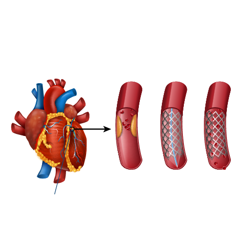
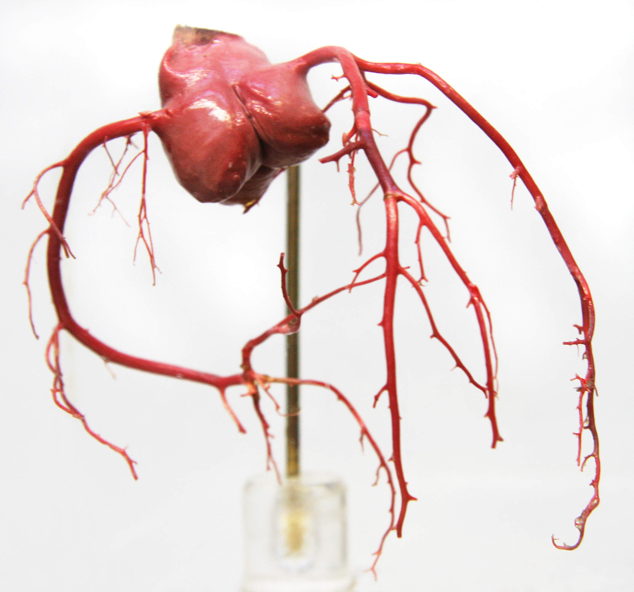
Coronary arteries are the vessels which supply blood (oxygen and nutrients) to the heart; this supply may be disrupted due to buildup of cholesterol and fatty deposits, called plaques, on the inner walls of the arteries (atherosclerosis) causing narrowing of the vessel lumen.

Coronary lesions branch out as they progress to supply various cardiac structures. A bifurcation lesion is a stenosis or abnormal narrowing of greater than 50% that occurs in a coronary artery at the origin of the side branch or adjacent to the origin of the side branch. Branching points a.k.a bifurcation points in the coronary arteries are prone to develop atherosclerosis as the shear stress is higher at the branch points.

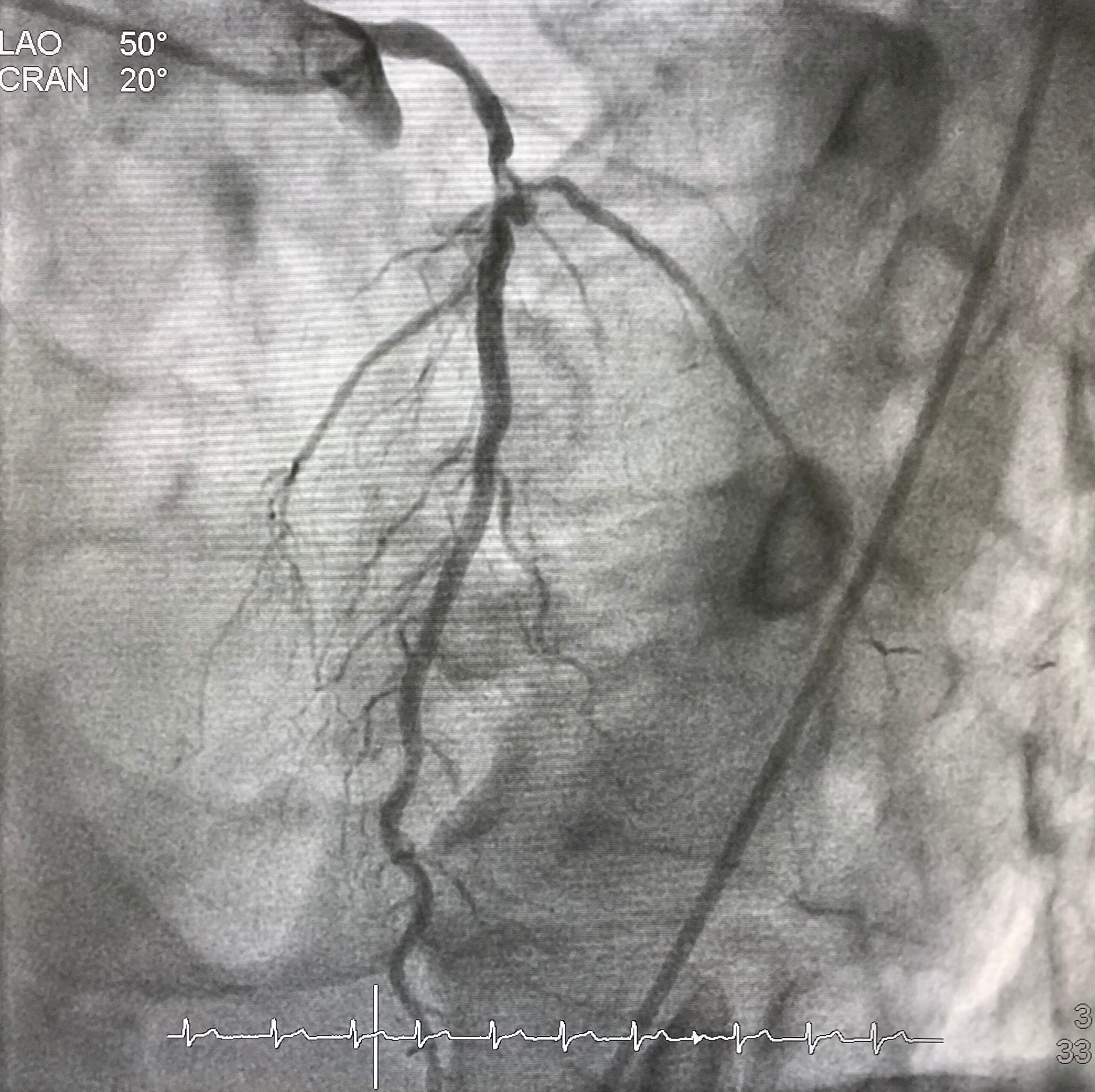
The figure shows how a bifurcation stenosis appears schematically as well as on angiography.

bifurcation lesion


Why bifurcation lesions are important?

A vast majority of coronary arteries obstructions happen at the branching point. About 15-20% of patients of referrals for bypass surgery are because of the presence of coronary narrowing at bifurcation location. They are considered a tough lesion to treat by angioplasty and stent.

How are bifurcation lesions diagnosed?

Bifurcation lesions are identified on coronary angiography. Only lesions affecting side branch blood vessels more than 2.25 mm is considered important and called bifurcation lesions. The main vessel that gives branches is called main vessel and the branches that originate from it are called side branches. The point at the division of the main vessel and side branch is called a bifurcation point.

How is a bifurcation lesion unique?

Bifurcation lesions can involve the coronary artery either before or after the bifurcation point. In addition, side branches could be either diseased or free from disease. Based on the location and severity of the cholesterol plaque at the bifurcation point, sub types of bifurcation lesions have been identified.

The challenge of performing angioplasty and stent for a bifurcation stenosis lies in the ability to preserve the main vessel and the side branch. Stents being metallic tubes risk occluding the side branch when placed across the side branch. This apprehension of losing the side branch, is one of the primary reasons for patients to be sent for bypass surgery instead of angioplasty and stent.

Coronary-bifurication

Can angioplasty and stent be performed for a bifurcation lesion?

Angioplasty and stent can be eminently performed for a bifurcation stenosis. Over the last 2 decades numerous techniques in stent implantation have been developed to treat the bifurcation lesions. Some of these techniques include:

  • Provisional T stent technique
  • Tap technique
  • Double kiss crush technique
  • Culotte technique

Advent of these techniques enabled experienced operators to perform angioplasty and stent successfully with chances of repeat narrowing seen in 3-6% patients at the site of stenting. Risk of losing a side branch after stenting is reduced to 1% once the new techniques are employed.

Dr. C Raghu is an expert in bifurcation lesion angioplasty and the center where he has been trained, ICPS Paris France is considered the “Mecca” for bifurcation lesion angioplasty techniques development. 

How does a bifurcation lesion angioplasty differ from a normal angioplasty?

Bifurcation lesion angioplasty, involves utilization of novel stent techniques mentioned above. In addition, angioplasty operator experience is a major factor in determining  outcomes. Choosing the “right” stent that allows side branch access, use of specialized technologies such as fractional flow reserve (FFR), expertise in deploying 2 stents especially the process of “stent crush” and the handling of two balloons through a single catheter – kissing balloon are key points for the best results in a stent procedure.

What are the chances of side branch occlusion in a bifurcation lesion angioplasty?

Contemporary bifurcation angioplasty using Intravascular imaging employing contemporary stenting techniques in the hands of expert bifurcation angioplasty operators carries a 99% success rate with a chance of losing side branch in 1% and repeat narrowing at the end of 9 months being 3-6%. These results are comparable and if not superior to bypass surgery as angioplasty entails a short hospital stay and fast recovery.

English Video

Book Online Consultaion


    Coronary Bifurcation Lesions Blog

    DOWNLOAD PDF

    Subscribe the Hearty Life Blogs


      Atrial Fibrillation


      Atrial fibrillation (AFib or AF) is when your heart’s upper chambers (Atria) beat irregularly and out of sync. The heart’s pumping function isn’t as strong or efficient as it…


      Atrial fibrillation (AFib) is one of the most common heart arrhythmias (irregular or abnormal beating). It happens when your heart’s upper chambers (the atria) beat…


      Atrial fibrillation, also known as afib or A-fib, is a medical condition that causes the heart to beat irregularly and quickly. It affects more than 3 million people in the US…


      Atrial fibrillation (AFib) interferes with the normal functioning of the heart. It can lead to blood clots, which, in turn, can result in a stroke or heart failure. That makes it…


      Atrial fibrillation is a progressive heart disease that can’t be cured. But the right course of treatment can help control afib symptoms and help patients live a close…


      Atrial Fibrillation is a chaotic, irregular beating of the heart. When the heart beating is irregular there will be stasis or stagnation of the blood within the heart chambers…


      What is Heart Failure? 

      Heart failure is not a single disease instead it is a constellation of symptoms. In this disease the heart  is unable to meet the requirements of the body by its inability to pump or be able to do so by increasing the filling pressures so that it might pump effectively.

      Heart Failure

      Heart Failure is not a disease but a  group of symptoms. It is the result of  many diseases affecting not only the  heart but other organs of body.

      Related : Types of Heart Failure

      Heart Failure vs Heart attack vs Cardiac Arrest

      All the three terms refer to different medical issues.

      • Heart failure is consequent to inefficient functioning of heart.
      • Heart attack is due to the interruption of blood supply to the heart.
      • Cardiac arrest is a situation where the heart stops to beat.

      Symptoms of Heart Failure

      Inefficient functioning of the heart leads to fluid logging in the body

      • Fluid logging in the lungs – presents as breathlessness – Left Heart failure in medical parlance Early stages of heart failure – breathlessness can be present on walking.
      • Advanced stages of heart failure –Breathlessness can be present at rest or, Inability to lie flat or may be awakened from sleep.
      • Fluid accumulation in other organs of the body – Right heart failure in medical parlance Legs causing swelling of the feet, Swelling of face, abdomen, Pain in upper right abdomen.
      • Heart failure symptoms which are related to the lungs are called left heart failure symptoms, those symptoms which are related to other organs are called right heart failure symptoms.
      • Concept of left and right heart
      • failure is important in treatment
      Related : What Is Systolic Heart Failure?

      What causes heart failure?

      Heart failure is a result of many disease processes in the body. Common among them being: Coronary artery disease

      • The most common disease that is responsible for heart failure is coronary artery disease. • Coronary artery disease means accumulation of cholesterol in the blood vessels supplying the heart leading to interruption of blood supply to the heart.This interruption of blood supply can either directly reduce the blood supply to the heart resulting in dysfunction or can cause heart attack which can also reduce the heart pumping ability.
      • Common diseases – such as diabetes, hypertension and sometimes abnormalities of the valves of the heart and fast beating of the heart of which most commonly atrial fibrillation can all result in reduced efficiency of the heart.

      In addition, advanced age itself beyond the age of 65-70 the efficiency of the heart reduces leading to heart failure.

      What tests are required for diagnosis of heart failure?

      The tests for the diagnosis of heart failure are

      • Imaging test
      • ECG
      • Blood test

      Blood tests in heart failure :

      • The most common blood test that is done is a pro-BNP test. If it is less than 125, we can safely exclude heart failure as the cause for breathlessness.
      • In addition, we look at presence of Anaemia, Renal dysfunction and blood glucose elevation in a patient with heart failure.

      Electrocardiogram (ECG) : This remains an important test

      Electrocardiogram (ECG) : This remains an important test

      ecg report

      • Can detect the presence of previous heart attack.
      • Irregular heart beat – most common is atrial fibrillation.
      • Presence of any Bundle branch block.

      Echocardiogram : The most common and important test for the diagnosis of heart failure is the  echocardiogram where we estimate the left ventricular ejection fraction (measures the pumping  ability of the heart) or in short called as EF.

      Other test done to manage heart failure are cardiac MRI and nuclear test.

      Related : Heart Failure – Role of Electrocardiogram (ECG)

      What is the role of Angiography in heart  failure?

      For every 3 patients with heart failure 2 have underlying obstruction to the heart’s blood supply. Identifying this is super important because removal of these obstructions by angioplasty and stent can lead to improved cardiac function. 

      The blood vessels supplying the heart are the  coronary arteries and obstructions are called coronary artery disease. So, whenever there is a  coronary artery disease, we can identify that only by performing a coronary angiography.

      The major advantage of this investigation is apart from diagnosis in coronary artery disease, in case  if any blocks in heart vessels are identified they can be treated by performing a balloon angioplasty  and a stent procedure.

      The benefit of removing the cholesterol plaques in the heart vessels is that – there can be a strong  chance for the heart functioning to recover once the blood flow is restored back to the heart.

      Book Online Consultaion


        Basics about Heart Failure Blog

        DOWNLOAD PDF

        Subscribe the Hearty Life Blogs


          Aortic-stenosis.jpg

          What is Aortic stenosis?

          Aortic stenosis is a disease where the valve between left ventricle and aorta is narrowed. Normally the left ventricle is the chamber which pumps blood to the entire body through the aortic valve. So if the aortic valve is narrowed either due to infection or age related degeneration it is called aortic stenosis. This is a disease of the elderly people usually beyond 50 years of age.

          What are the symptoms of aortic stenosis?

          When the disease is severity is mild people usually do not experience any symptoms. But is the disease is severe – even though the disease is severe in early phases the patient might not have symptoms. 

          So, early stages of aortic stenosis patient might not have any symptoms. Where as in the advanced stage of severe aortic stenosis the patients can develop can develop chest pain also called angina in medical terminology. So chest pain which increases on walking or any other form of exertion and gets relieved on rest or stopping of that activity is called angina. So patients with aortic stenosis because is unable to pump as per the requirements of the body (due to aortic valve narrowing) they experience angina. 

          The other symptom is breathlessness – on walking or on lying down flat the patient develops difficulty in breathing. This is referred to as dyspnea in medical terminology. So when ever a patient is having a fixed blood supply to various organs without increasing as per their needs there is pooling of blood in the heart. This pooled blood in the heart “reverses back” into the lungs which is responsible of breathlessness.

          Finally in advanced stages of aortic stenosis patients develop a sudden loss of consciousness with spontaneous recovery. These episodes of loss of consciousness are also called as syncope in medical terminology.

          So the predominant symptoms of aortic stenosis are chest pain, breathlessness and sudden loss of consciousness.

          At the same time patients with aortic stenosis will develop an impaired function of the heart also called heart failure. This heart failure need to necessarily present in severe heart failure but can also be seen in intermediate or moderately severe aortic stenosis. 

          Aortic stenosis and high blood pressure 

          Patients with aortic stenosis have reduced supply of blood to various organs of the body. Because of this it was believed that patients with aortic stenosis tend to have low blood pressure. This is not true regarding the current epidemic of aortic stenosis we are currently seeing. Currently most of the aortic stenosis patients are elderly in their 60s, 70s and 80s of age. So these patients because of the progression of the age and hardening of the blood vessels they develop high blood pressure or Hypertension in medical terminology. So patients with aortic stenosis are not spared from high blood pressure contrary to what we were believing till date and what we are seeing is a scenario of aortic stenosis patients having high blood pressure levels.

          What is the impact of this high blood pressure on a patient with severe aortic stenosis?

          Patients with high blood pressure and severe aortic stenosis develop a faster progression of the disease severity. So a patient of aortic stenosis with uncontrolled blood pressure can have a severe aortic stenosis at a much earlier age. 

          How can patients with aortic stenosis control their blood pressure?

          People with aortic stenosis and high blood pressure need to control their blood pressure using 3-4 different types of medicines. A good control of blood pressure is one of the first steps in retarding the progression of aortic stenosis. 

          Can people with aortic stenosis do exercise? 

          People with aortic stenosis tend to have a fixed cardiac output. This means – the aortic valve is narrowed and this narrowing limits the blood supply to various organs of the body. When there is a reduction in blood supply to various organs of the body – the first to be affected is the brain. This causes syncope or sudden loss of consciousness. 

          Exercise AS

          So when a person with severe aortic stenosis exercises vigorously then there is a reduction of blood supply to the brain causing sudden unconsciousness. This problem happens in people with an advanced or severe aortic stenosis. So people with severe or advanced aortic stenosis are advised not to participate in vigorous physical activity such as running, jogging or weight lifting etc. 

          But at the same time as we all know for the control of BP, blood sugar and cholesterol and effective control of heart failure are important steps for retarding the progression of aortic stenosis. So a mild to moderate severe intensity exercise is advised for control of the various co-morbidities in aortic stenosis. But at the same time a vigorous or severe intensity exercise is definitely not to be performed. Severe intensity exercise or competitive sports is a contra indication for aortic stenosis patients in medical terminology.

          Aortic stenosis

          Aortic Stenosis Symptoms (Telugu)

          Book Online Consultaion


            Aortic Stenosis : Symptoms Blog

            DOWNLOAD PDF

            Subscribe the Hearty Life Blogs


              Cong.-Heart-Disease.jpg

              Congenital heart diseases are diseases that are present from birth and affect the normal heart functioning. These are the most frequently occurring congenital disorder, responsible for 28% of all congenital birth defects. The birth prevalence of CHD is reported to be 8-12/1000 live births. Considering a rate of 9/1000, about 1.35 million babies are born with CHD each year globally.

              What are the Causes of Congenital Heart Defects?

              In most cases, no obvious cause of congenital heart disease is identified. However, some things are known to increase the risk of the condition, including:

              • Down’s syndrome – a genetic disorder that affects a baby’s normal physical development and causes learning difficulties
              • the mother having certain infections, such as rubella, during pregnancy
              • the mother taking certain types of medicine during pregnancy, including statins and some acne medicines
              • the mother smoking or drinking alcohol during pregnancy
              • the mother having poorly controlled type 1 diabetes or type 2 diabetes
              • other chromosome defects, where genes may be altered from normal and can be inherited (run in the family)

              Many cases of congenital heart disease can be diagnosed before a baby is born during an ultrasound scan in pregnancy. At times a focused foetal heart scan called foetal echocardiography in specialized centers helps to diagnose the cardiac problem before birth. However, it’s not always possible to detect congenital heart defects in this way.

              congenital heart disease

              Signs and Symptoms of Congenital Heart Disease

              Congenital heart disease can have a number of symptoms, particularly in babies and children, including:

              • Rapid heartbeat
              • Rapid breathing
              • Swelling of the legs, tummy or around the eyes
              • Extreme tiredness and fatigue
              • A blue tinge to the skin or lips (cyanosis)
              • Tiredness and rapid breathing when a baby is feeding

              These problems are sometimes noticeable soon after birth, although mild defects may not cause any problems until later in life.

              Types of Congenital Heart Disease

              There are many types of congenital heart disease and they sometimes occur in combination. Some of the more common defects include:

              • Septal defects – where there’s a hole between 2 of the heart’s chambers (commonly referred to as a “hole in the heart”)
              • Coarctation of the aorta – where the main large artery of the body, called the aorta, is narrower than normal
              • Pulmonary valve stenosis – where the pulmonary valve, which controls the flow of blood out of the lower right chamber of the heart to the lungs, is narrower than normal
              • Transposition of the great arteries – where the pulmonary and aortic valves and the arteries they’re connected to have swapped positions
              • Underdeveloped heart – where part of the heart doesn’t develop properly making it difficult for it to pump enough blood around the body or lungs.

              congenital heart disease

              Treating Congenital Heart Disease

              Treatment for congenital heart disease usually depends on the defect you or your child has.

              Mild defects, such as holes in the heart, often don’t need to be treated, as they may improve on their own and may not cause any further problems.

              Surgery or interventional procedures are usually required if the defect is significant and causing problems. Modern surgical techniques can often restore most or all of the heart’s normal function.

              However, people with congenital heart disease often need treatment throughout their life and therefore require specialist review during childhood and adulthood. This is because people with complex heart problems can develop further problems with their heart rhythm or valves over time.

              Most surgery and interventional procedures aren’t considered to be a cure. The affected person’s ability to exercise may be limited and they may need to take extra steps to protect themselves from getting infections.

              It’s important that a person with heart disease and their parents or carers discuss these issues with their specialist medical team.

              Book Online Consultaion


                Basics of Congenital Heart Disease Blog

                DOWNLOAD PDF

                Subscribe the Hearty Life Blogs

                  dr. raghu

                  DR. RAGHU | Best Cardiologist in Hyderabad

                  MD, DM, FESC, FACC, FSCAI

                  Cardiology Coronary, Vascular and

                  Structural Interventions


                  Conditions & Diseases

                  Coronary angioplasty

                  Angioplasty

                  Coronary angioplasty

                  Aortic Stenosis

                  Coronary angioplasty

                  Atrial Fibrillation

                  Coronary angioplasty

                  Atrial Septal Defect


                  View More Services


                  WhatsApp-Image-2021-11-01-at-3.35.00-PM-1200x683.jpeg

                  Women especially at young age are experiencing an unprecedented increase in heart attacks and strokes. We attempt In this article to make the reader aware of the reasons for this.

                  How are women different from men for developing heart attack?

                  Women tend to develop cardiac disease 7-10 years later compared to men that is attributed to the protective effect of estrogen hormone, which is present till menopause.

                  What are the traditional risk factors for developing heart disease?

                  Both men and women are prone to develop cardiac problems if they have one or multiple risk factors listed below. They are called traditional because they have been in practice for the past 6 decades.

                  Non-modifiable risk factors Modifiable risk factors
                  ·         Age

                  ·         Male gender

                  ·         Family history of cardiac disease (< 60 years)

                  ·         Smoking

                  ·         Diabetes

                  ·         Hypertension

                  ·         Hyperlipidemia

                  ·         Obesity

                   Despite the absence of many of these risk factors, women worldwide are increasingly dying due to cardiac ailments.

                  Do traditional risk factors confer a disparate risk for developing cardiac problems in women?

                  Women are sensitive than men for the development of heart diseases when they have associated traditional risk factors.

                  For example:

                  • Diabetes: Women with diabetes have a 7-fold increase in cardiac events compared to only 3-fold increase in diabetic men.
                  • Smoking and tobacco use has been shown to enhance the risk by an additional 25% in women compared to men for development of heart attacks. In fact tobacco use has been responsible for 50% of cardiac events in women and confers a 3-fold increased events. Normally women develop heart attacks 7-10 years later than men due to the protective benefits of estrogen hormone. This protective effect is lost with tobacco use and if they develop diabetes.

                  Unique risk factors for cardiac problems in female gender include: 

                  • Pre-eclampsia (High BP during pregnancy associated with complications)
                  • Diabetes during pregnancy
                  • Polycystic Ovary syndrome
                  • Early menopause
                  • Autoimmune diseases

                  Early menopause and risk of heart attack:

                   About 10% of women experience menopause naturally before the age of 45 years. This is called early menopause. Because of lack of estrogen in post menopausal women, it predisposes to cardiac events.

                  Compared to women of similar age, those who attain natural or surgical menopause (removal of uterus – Hysterectomy) less than 45 years are at 50% higher risk of developing heart attack and 20% increased risk of death.  Those women who attained surgical menopause tend to have higher cardiac events compared to natural menopause. So women should be dissuaded to undergo uterus removal surgery unless there is a clear evidence that the procedure is definitely required. Hormone replacement therapy has been tried to mitigate this risk but in vain. Current guidelines are against hormone replacement therapy for early menopause.

                  What can we do to prevent these cardiac events in women?

                  Targeting traditional risk factors – diabetes, hypertension, lipids, sedentary life styles, inappropriate food choices and obesity form the bedrock strategy for reducing the risk of developing heart attacks.

                  But for women further measures are needed to address the unique risk factors for women. The American College of Cardiology in its 2019 guidelines notified pre eclampsia, early menopause and autoimmune disease as “risk enhancers” for cardiac disease. They also added that physicians should have a low threshold to treat those patients with risk enhancers by life style measures and cholesterol lowering drugs to reduce the risk of developing heart attacks.

                  Finally women should realize that they are no longer at low risk for developing cardiac ailments and need to understand there are new risk factors that put them at “enhanced risk” for developing heart attacks.


                  mitral-balloon.jpg

                  Mitral stenosis is the narrowing of the valve present between the left chambers of the heart (mitral valves), thereby blocking the blood flow. This condition usually develops several years after a person had a rheumatic fever. Mitral stenosis is treated with balloon valvotomy, when the medications do not reduce the symptoms. If left untreated, mitral stenosis can result in various heart complications.

                  Mitral valvotomy (or valvuloplasty), also known as percutaneous balloon dilation, is a minimally invasive procedure that involves widening a mitral valve using a balloon catheter, a thin, flexible tube with a balloon at the tip. This procedure improves the overall function of the heart.

                  What are the risks of mitral valvuloplasty?

                  The risks associated with percutaneous balloon dilation include:

                  • Blood clot formation or tears in the heart
                  • Backward flow of the blood (mitral valve regurgitation) due to damaged valve
                  • Restenosis of the mitral valve

                  What happens before the procedure?

                  Your doctor will explain the procedure in detail and provide you the opportunity to ask any questions; do not hesitate to ask any questions related to the procedure.

                  Your healthcare team will give you certain instructions to prepare for the procedure:

                  • You will be asked not to eat or drink anything after midnight, on the previous night of the procedure; you can drink water up to 4 hours of the procedure.
                  • A blood test may be done to evaluate the time required for the blood to clot; other blood tests may also be performed.
                  • You may be asked to stop taking certain medicines that may involve with the blood clotting process.

                  Notify your doctor if you:

                  • Had an allergic reaction to any contrast dye, iodine or seafood.
                  • Are sensitive to any medications, tape, latex, or anaesthetic agents.
                  • Have a pacemaker.
                  • Have any body piercings on the abdomen or chest.
                  • All the medications you are taking, including over-the-counter drugs, herbal supplements, blood thinners, etc.
                  • Have heart valve disease, as antibiotic drugs may be given before this procedure.
                  • Have a history of bleeding disorders
                  • Are or may be pregnant

                  Before the procedure, the area near the catheter insertion site (the groin area) may be shaved. Your physician may order other preparations for the procedure based on your medical condition.

                  How is mitral valvuloplasty performed?

                  On the day of the procedure, you should remove your jewellery and other objects that may interfere with the procedure. You will be asked to change into a hospital gown and empty your bladder before the procedure.

                  An intravenous (IV) line will be attached to your arm or hand to inject medications and administer IV fluids, if needed. You may receive a sedative to help you relax.

                  The following are the steps generally involved in a balloon valvuloplasty:

                  • A local anaesthetic is injected at the insertion site.
                  • Once the anaesthesia sets in, a sheath or an introducer (a plastic tube) will be inserted into the blood vessel.
                  • A valvuloplasty catheter is inserted through the sheath into the blood vessel. The catheter is advanced through the aorta into the heart valve. The catheter may be guided by a fluoroscopy (a special x-ray).
                  • Once the catheter reaches the precise location, a contrast dye is injected into the valve to get a clear image of the area.
                  • The balloon is then inflated and deflated several times.
                  • The catheter is then removed.
                  • The catheter insertion site is closed using a sterile bandage.

                  What happens after the procedure?

                  After the procedure, you will be moved to the recovery room. You should remain flat on bed for several hours after the procedure. Your vital signs, and circulation and sensation in the affected arm or leg, the insertion site will be monitored regularly.

                  Medicines may be given for pain or discomfort near the insertion site. You will be asked to drink water and other fluids to eliminate the contrast dye from your body. You can return to your regular diet after the procedure.

                  Mostly, you may have to spend the might in the hospital, based on your medical condition and recovery.

                  Your healthcare team will give you instructions to be followed after leaving the hospital:

                  • Keep a check on the insertion site for unusual pain, bleeding, swelling, or discoloration.
                  • Keep the insertion site clean and dry.
                  • Do not participate in any strenuous activities. Your doctor will inform you when you can resume normal activities and return to work.

                  Call your doctor immediately if you have any of the following:

                  • Fever or chills
                  • Severe pain, swelling, redness, bleeding or other leakage from the insertion site
                  • Numbness, coolness or tingling sensations in the affected extremity
                  • Pain or pressure in the chest, nausea or vomiting, sweating, or dizziness
                  • Reduced urination
                  • Swelling of the abdomen or extremities
                  • Over 3 pounds weight gained in a day


                  diagnosis-procedure-carotid_and_cerebral.jpg

                  Carotid artery disease is a condition that arises when the fat deposits accumulate and block the blood vessels that deliver blood to the brain. People with carotid artery disease are at higher risk of developing stroke. So, to determine the risk and prevent the associated complications, a procedure known as cerebral angiography is recommended.

                  Cerebral angiography is a diagnostic procedure that uses X-rays to evaluate the blockage or any brain abnormalities in the carotid arteries (blood vessels in the brain and neck).

                  When is cerebral angiography recommended?

                  Cerebral angiography is recommended if the doctor suspects any of the following abnormalities within the brain:

                  • An aneurysm
                  • A dilated blood vessel in the brain
                  • Brain tumour
                  • Brain clot
                  • Stroke

                  It is also used to evaluate arteries in the head and neck before undergoing any surgery to provide additional information regarding any abnormalities that are not visible on other imaging tests, and as a minimally invasive procedure to treat vessel abnormalities.

                  In some cases, it is done to determine the underlying cause of the following symptoms:

                  • Severe headache
                  • Memory loss
                  • Dizziness
                  • Blurred vision
                  • Loss of balance or coordination
                  • Weakness or numbness

                  Before the procedure:

                  Specific instructions are given before undergoing the procedure. These instructions may include:.

                  • Any allergies
                  • The use of current medicines, vitamins, and mineral supplement
                  • The current medical conditions
                  • Any food or dietary restrictions before the procedure.

                  What happens in the procedure?

                  Before initiating the procedure, the patient’s head is stabilized by using a strap, tape, or sandbags. Based on the age of the person, either local or general anaesthesia, is administered. Once the anaesthesia sets, the doctor will sterilize the groin region and make an incision. Under the guidance of X-rays, the catheter is passed into an artery in the neck.

                  Once the catheter is placed in the correct position, the contrast dye is injected to highlight the blockage. After the X-rays are done, the catheter is removed, and the incision is closed.

                  What to expect after the procedure?

                  Once the procedure is done, the vitals are monitored in a recovery room. The person would be instructed to keep the leg straight if the catheter is administered through the groin region. Pain and inflammation at the site of incision are common, which can be relieved by applying ice packs and taking the prescribed medicines.

                  What considerations should be taken after the test?

                  Cerebral angiography is a diagnostic procedure, so there are not much considerations. However, following the below-given tips may ease the discomfort associated with the test:

                  • Have a healthy and well-balanced diet.
                  • Do not lift heavy weight for a few days or as suggested by the doctor.
                  • Perform normal activities 8 to 12 hours after the procedure.

                  Talk to the doctor:

                  Call the doctor immediately on noticing any of the following symptoms:

                  • Chest pain
                  • Dizziness
                  • Infection at the catheter site
                  • Shortness of breath
                  • Skin rash
                  • Slurred speech
                  • Vision problems
                  • Numbness in the face, arms, or leg muscles

                  Book Online Consultaion


                    Carotid and Cerebral angiography Blog

                    DOWNLOAD PDF

                    Subscribe the Hearty Life Blogs


                      cci-dialysis_fistuloplasty.jpg

                      Hemodialysis is a preferred treatment option for patients with chronic kidney disease. In this t procedure, the blood is filtered outside the body by a dialyzer or “artificial kidney”. For hemodialysis, a vascular access is created to insert the needles that connect the dialyzer, thereby allowing the blood to move out and return to the body at a high rate. An arteriovenous fistula is the preferred vascular access for hemodialysis.

                      An arteriovenous (AV) fistula is created by connecting an artery to a vein, usually in the wrist or upper arm. But sometimes, the fistula can become infected, blocked or narrowed. The blocked fistula can be treated by a balloon fistuloplasty.

                      What is balloon fistuloplasty?

                      Balloon fistuloplasty is a procedure in which any blockage or narrowing in the fistula is located by using a dye, and the blockage is relieved by stretching the blood vessels with a special balloon.

                      In this procedure, a small balloon is inflated for several times at the narrowed regions of a fistula; if required a stent may also be placed. This technique widens the lumen and facilitates the process of dialysis.

                      Why perform fistuloplasty?

                      A fistula can age and cause problems like clotting and scarring, thereby decreasing its function and effectiveness of dialysis. Clots can decrease the rate of blood flow or block the fistula completely. If these problems are left untreated, it may lead to the failure of fistula.

                      Thus, it is important to treat the narrowing or blockage at an early stage, so that the fistula works well, and dialysis occurs without any complications.

                      What are the risks of dialysis fistuloplasty?

                      Fistuloplasty is usually a safe procedure, but some complications may occur rarely. The common risks and complications associated with fistuloplasty include:

                      • Bruising around the site of insertion of the needle, which may become large and uncomfortable (rarely).
                      • Infection of large bruises, which may need antibiotic treatment or surgical intervention.
                      • Allergic reaction to the dye, which may present as a skin rash
                      • Circulatory problems due to the damage to the artery or fistula caused by catheter or balloon
                      • Treatment failure, which will require surgical intervention

                      How to prepare for the procedure?

                      Your healthcare provider will give you instructions to prepare for the procedure, which may include:

                      • You may be asked not to eat or drink anything for six hours before the procedure; you can drink water up to two hours before.
                      • Some blood tests may be performed before the procedure to evaluate the risk of bleeding.
                      • If you are diabetic, ask your doctor to alter the treatment regimen.
                      • Ask your doctor which medicines you can continue to take, and which ones you should stop.
                      • If you are taking any antiplatelets or anticoagulants, you may have to stop taking these medicines a few days before.
                      • Ensure that you have an adult to drive you home and accompany you overnight.

                      Inform your healthcare provider if you:

                      • Are allergic to iodine, or have any other allergies
                      • Have a history of reaction to the dye used for CT scan or X-rays
                      • May be or are pregnant
                      • Are a diabetic

                      What happens during the procedure?

                      The following are the steps usually performed in a fistuloplasty:

                      • You will be asked to lie on your back on an x-ray table. Some monitoring equipment will be attached to measure your heart rate and blood pressure.
                      • The interventional radiologist will observe the fistula by using an ultrasound, which provides a clear picture of the fistula.
                      • A small needle is inserted in the fistula, and a contrast dye is injected. This provides an image of the blood vessels, which helps to locate the narrowing or blockage.
                      • A catheter (a small, flexible tube) with a balloon at the tip is then inserted in the blood vessel to reach the precise location.
                      • Once the catheter reaches the site of the blockage or narrowing, the balloon is inflated and deflated several times from outside the body.
                      • Sometimes, if the balloon does not improve the fistula, a permanent stent may be used to widen the narrowing.
                      • Then the catheter is removed from the blood vessel, and the puncture site is stitched to prevent bleeding.

                      Usually, fistuloplasty takes about one hour, but the duration may vary in different patients.

                      What happens after the procedure?

                      You will have to stay in the hospital after the procedure for three to four hours for observation. Your pulse, blood pressure and oxygen levels in the body will be monitored regularly. You may return to your normal diet. The fistula is ready to use immediately after the procedure.

                      Your nurse will tell you when you can go home. You will need a friend or a family member to drive you home; using public transport is not recommended.

                      Your fistula should be ready for use immediately after the procedure.

                      What measures do I take after going home?

                      The following measures will help you recover better after a fistuloplasty:

                      • Rest well on the day of the procedure and the next day. Then, you can return to your normal activities.
                      • You can follow your normal diet.
                      • Take the pain killers as prescribed r instructed.
                      • Continue to take you regular medicines, as prescribed.
                      • Do not take metformin until two days after the procedure.
                      • You can have a bath or shower the next day.

                      Call your doctor immediately

                      if you have any of the following symptoms:

                      • A lot of swelling and bruising
                      • Severe pain at the puncture site that does not get better with painkillers
                      • Bleeding at the puncture site
                      • Change in the colour of your arm
                      • Fever or chills
                      • A lump, pus or discharge at the puncture site
                      • Difficulty breathing or chest pain


                      diagnosis-procedure-cardiac_catheterization.jpg

                      Cardiac catheterization is one of the most extensively performed cardiac procedures. If you have chest pain or irregular heartbeat, your doctor may recommend cardiac catheterization.

                      Cardiac catheterization is a minimally invasive procedure used to detect and treat cardiovascular diseases. This procedure involves the insertion of a catheter (a thin hollow tube) into the large arteries or veins present in the neck, arm or groin, which is then guided to the heart using a special X-ray. Once the catheter reaches the location, diagnostic tests or treatment procedures are carried out.

                      Why perform Cardiac Catheterization?

                      Cardiac catheterization is done to find the cause of the signs and symptoms of a heart problem or to treat or repair the heart damage.

                      This procedure is performed to:

                      • Determine the pressure levels in the chambers of the heart.
                      • Determine the heart function after a cardiac intervention.
                      • Detect blockages in the coronary arteries (coronary angiography) or valve dysfunction.
                      • Perform procedures such as angioplasty, ablation therapy or valve repair.
                      • Obtain a small piece of heart tissue to examine under a microscope for detecting conditions affecting the heart muscle (cardiac biopsy).

                      What is the difference between Cardiac Catheterization and Coronary Angiography?

                      Catheterization is a procedure used to diagnose and treat various conditions. It involves the insertion of a catheter into the arteries and veins to reach the heart and to measure how much blood the heart pumps out, the pressure in each heart chamber, and to detect any defects in the heart. Also, various small instruments can be inserted into the catheter to view the interior of the blood vessels, to remove a tissue sample from the heart for further examination, etc.

                      Coronary angiography is a type of catheterization procedure, which involves analysing the arteries that supply the heart with oxygen-rich blood. This procedure involves the insertion of a catheter in the arm or the groin, which is threaded to reach the coronary arteries. During insertion, fluoroscopy (a continuous x-ray procedure) is used to guide the catheter to reach the precise position. After the catheter tip is at the right location, a contrast dye is injected into the coronary arteries. This dye can be seen on x-rays, and the outline of the arteries is viewed on a video screen.

                      How do I prepare for the procedure?

                      Inform your doctor about:

                      • The medications that you are taking, especially blood-thinners or anti-inflammatory medications
                      • Any medical conditions that you have
                      • Allergies to any dyes or specific medications

                      Before the procedure:

                      • Fast for at least 6 hours, as food and liquid increase the possibility of complications with general anaesthesia.
                      • Empty your bladder before heading to the procedure.
                      • Remove dentures and jewellery, as they may interfere with the imaging procedures.
                      • Your general health status and vital signs will be monitored.

                      What happens during the procedure?

                      During cardiac catheterization, an IV cannula is inserted in your arm to administer medications. You will be given a mild sedative to help you relax during the procedure.

                      The general procedure of cardiac catheterization is given below:

                      • The site of catheter insertion (usually the groin) is cleaned and shaved.
                      • Usually, local anaesthesia is administered. General anaesthesia may be given before surgical procedures such as valve repair or ablation.
                      • When the site of catheter insertion becomes numb, an incision is made, and the catheter is inserted using a catheter sheath.
                      • Once the catheter reaches the heart, a dye is administered which outlines the vessels, valves and chambers of the heart.
                      • The diagnostic tests or surgical procedures will then be performed for treating the condition.

                      Vital signs and electrocardiogram will be continuously monitored during the procedure.

                      Cardiac catheterization usually lasts for about 30 minutes. It may be longer if you are undergoing any other test or intervention using this procedure.

                      After the procedure is done, the catheter will be removed, and the incision will be closed by stitches. A sterile dressing will be used to prevent infection in the incision site.

                      What happens after the procedure?

                      Once the procedure is done, you will be asked to lie flat on the bed for 2-6 hours after the procedure. Your heart rate and blood pressure will be continuously monitored.

                      Once the anaesthesia wears off, you will be given pain medications. You will be asked to drink plenty of water to eliminate the dye that was administered. Usually, you will be asked to stay in the hospital for a few hours or a day.

                      After leaving the hospital, follow all the post-procedural instructions given by your doctor. You may resume your normal activities in a day’s time. Some soreness at the incision site is normal; it may reduce within a week.

                      What are the risks of Cardiac Catheterization?

                      Cardiac catheterization is a relatively safe procedure. However, like any other invasive procedure, some uncommon risks of this procedure are:

                      • Infection
                      • Blood clotting
                      • Bruising at the incision site
                      • Allergic reaction to dye
                      • Arrhythmia
                      • Air embolism
                      • Stroke

                      Call your doctor if you experience:

                      • Severe pain at the incision site
                      • Signs of infection: redness, warmth, pus oozing or excessive swelling around the incision site
                      • Numbness or tingling sensation in your limbs


                      +91 95424 75650

                      Call us now if you are in a medical emergency need, we will reply swiftly and provide you with a medical aid.


                      +91 95424 75650


                      Call us now if you are in a medical emergency need, we will reply swiftly and provide you with a medical aid.


                      Dr. Raghu | Heart Specialist in Hyderabad
                      Yashoda Hospitals, Sardar Patel Rd, behind Hari Hara Kala Bhavan, Kummari Guda, Shivaji Nagar, Secunderabad, Telangana 500003


                      Copyright © 2023, Dr. Raghu. All rights reserved.

                      [gtranslate]