Resources / Dr Raghu

cci-cardiac_catheterization.jpg

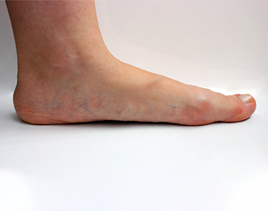
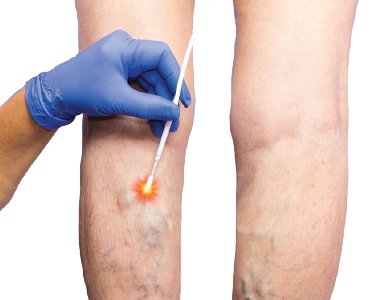
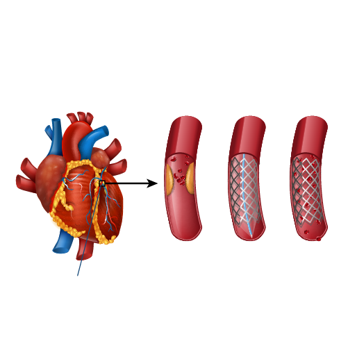
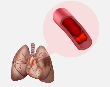
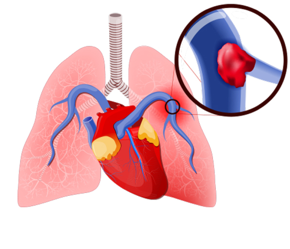
Peripheral arteries are the blood vessels that deliver blood to the lower limbs. When the cholesterol accumulates in these blood vessels, the blood flow to the lower limbs gets blocked. Peripheral angioplasty is a minimally invasive procedure. It is done to restore the blood flow by opening the blocked peripheral arteries.

When is peripheral angioplasty performed?

  • Peripheral angioplasty is mostly indicated in the treatment of Peripheral Vascular Disease (the circulatory disorder caused by the blocked or narrowed blood vessels outside the heart).
  • Peripheral artery disease is the common condition, in which peripheral angioplasty is recommended.

Are there any risks with peripheral angioplasty?

Peripheral angioplasty is associated with the following risks:

  • Breathing problems
  • Bleeding
  • Blood clots
  • Infection
  • Kidney damage
  • Damage to the blood vessel

How to prepare for the procedure?

Before initiating the procedure, a physical examination and imaging tests are done to determine the overall health condition. Additionally, the following steps would help in a successful procedure and quicker recovery:

  • Inform the use of current medications, vitamin or mineral supplements.
  • Tell the doctor about any underlying disease condition.
  • Take the prescribed medicines.
  • Notify the doctor if you have any allergies.
  • Six to eight hours before the surgery do not eat or drink anything.

What happens during the procedure?

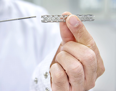
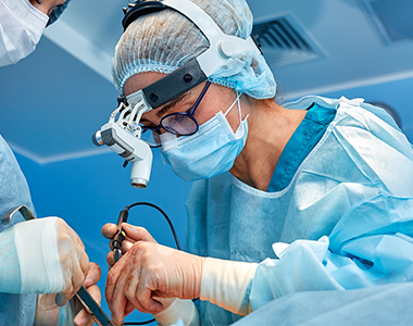
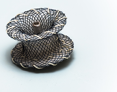
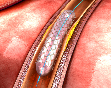
Local anaesthesia is administered in the upper thigh region. Once the anaesthesia sets, an incision is made on the upper thigh to insert the catheter. By using a high-resolution fluoroscopic, the catheter is guided to the blocked artery. When the catheter reaches the obstructed artery, the balloon is inflated to widen the blood vessel. Once the blood flow is restored, a stent is placed to prevent the risk of further blockage. Finally, the incision is closed and covered with a sterilized bandage.

What to expect after the procedure?

You will be placed in a recovery room and the vital parameters would be checked. For at least 3-6 hours, you need to remain still, to prevent bleeding from the incision site. Depending on the patient’s condition, the doctor will decide whether the person requires a hospital stay or not.
Before discharge, you will receive the following instructions:

  • Keep the wound clean and dry.
  • Do not lift heavyweights.
  • Avoid strenuous exercises for at least 24 hours after the procedure.
  • Drink plenty of fluids to help flush out the contrast dye from the body.

Care after peripheral angioplasty:

Although peripheral angioplasty clears the blockage, it does not treat the underlying cause of the blockage. So, to prevent the further risk of blockage, the following steps should be taken:

  • Maintain a healthy body weight.
  • Quit smoking.
  • Exercise regularly.
  • Take the prescribed medicines to prevent re-narrowing of the blood vessels.
  • Manage stress.
  • Avoid fatty-foods.
  • Have a low-salt and low-fat diet to prevent the risk of fluid retention.

Seek medical attention:

The following symptoms are the warning signs that require immediate medical attention:

  • Swelling in limbs
  • Chest pain
  • Shortness of breath
  • Fever associated with chills (over 101oF)
  • Weakness
  • Dizziness


disease-condtions-critical_limb_ischemia.png

Critical limb ischemia (CLI) refers to severe compromise of blood flow to alimb (hands or legs) which causes severe limb pain at rest or even loss of limb. It is the most advanced form of peripheral artery disease.

CLI numbers:

  • Prevalent in 2% patient over 70 years of age.
  • Within 1 year of diagnosis, 40-50% patientshave an amputation and 25% die.

How CLI leads to amputation?

Amputation occurs when there is marked ischemia of the limb owing to reduction of blood flow and increase demand in the limb.

Factors that reduce blood supply:

  • Diabetes mellitus
  • Severe Renal or Heart failure
  • Vasospastic diseases
  • Smoking

Factors that increase demand for blood flow:

  • Infection (cellulitis)
  • Skin breakdown
  • Trauma
  • Osteomyelitis

 Diagnosis of CLI:

Characteristic Duplex Ultrasound Digital – Subtraction Angiography Magnetic Resonance Angiography (MRA) Computed Tomographic Angiography (CTA)
Advantages
  • Noninvasive
  • Can Visualize & Quantitate severity
  • Gold Standard
  • High Resolution
  • Can guide intervention
  • Noninvasive
  • No radiation
  • No contrast
  • 3 D
  • Noninvasive
  • Higher Resolution than MRA
  • 3 D
Disadvantages
  • Operator dependent
  • Limited by
    dense calcification
  • Invasive
  • Radiation
  • Contrast
  • 2Dimensional
  • Lower Resolution than CTA
  • Claustrophobia
  • Contrast Image artifact if stent present
  • Radiation (25% of dose with DSA)
  • Contrast
  • Limited by calcification

Management plan for critical limb ischemia:

Critical Limb Ischemia Non-Healing Ulcer Rest Pain

MRA, CTA or invasive argiography shows lesion treatable by endovascular approach

  • Endovascular revascularization ->Wound care and atherosclerosis risk factor modification
  • Lesion treatable by open sugery at acceptable operative risk
    • Surgical revascularization ->Wound care and atherosclerosis risk factor modification
    • Consider primary amputation ->Wound care and atherosclerosis risk factor modification


cci-carotid_artery_stenting.jpg

Carotid arteries are the major blood vessels on each side of the neck that supply blood to the brain, face and neck. These arteries extend from the aorta in the chest to the base of the skull.

Over time, the arteries can harden and cause a build-up of plaque (calcium, cholesterol, and fibrous tissue deposits) on the walls of the arteries. This plaque build-up can narrow and stiffen the arteries. The progressive plaque build-up can reduce the blood flow through the arteries or cause the formation of blood clots. Such narrowed carotid arteries can be treated by using stents.

Carotid artery stenting (CAS) involves the insertion of a metal-mesh tube, called a stent, at the site of clogged arteries, to expand the lumen of the arteries and increase the blood flow to the brain.

What are the indications and contraindications for CAS?

The indications for CAS include:

  • High risk of stroke
  • Carotid artery blockage of 70 percent or more
  • Intolerance to general anaesthesia for carotid endarterectomy (an open surgery to remove the plaques in carotid arteries and to reduce the risk of stroke)
  • Damage to the contralateral vocal cord caused by previous carotid endarterectomy or neck surgery
  • Narrowing of carotid artery after previous CEA
  • Neck irradiation

The contraindications for CAS are:

  • Allergic reaction to intravenous (IV) contrast dye in the past
  • Unstable carotid or aortic arch plaque
  • A recent stroke in less than 14 days
  • Total thrombotic occlusion of carotid artery

What are the complications of CAS?

Here are some complications that may occur during or after CAS:

  • An embolism, blockage due to a clot or debris in an artery in the brain, a serious complication which can cause a stroke.
  • Formation of a blood clot along the stent or a tear in the artery wall (dissection).
  • Restenosis, the blockage of the carotid artery after the procedure.
  • Kidney damage, particularly in individuals with kidney problems, caused by the dye used for angiogram.
  • Bleeding from the incision site in the arm or groin artery, known as false aneurysm or hematoma (an unusual complication).
  • Mild tenderness and bruising at the puncture site, which usually resolves over time.

The factors that increase the chance of complications during CAS include:

  • Age > 80 years
  • High blood pressure
  • Allergy to contrast material
  • Sharp bends or other structural abnormalities in the carotid arteries
  • Significant atherosclerosis or plaque build-up in or near the carotid artery
  • Widespread blockages in the arteries in the legs and arms
  • Poor kidney function

How to prepare for CAS?

When planning for the procedure, inform your doctor about:

  • All the medicines that you take, including over-the-counter medicines, blood thinners, herbs, supplements, etc.
  • Habits like smoking; your doctor may help you quit smoking
  • Any changes in your health, like a fever
  • Are or may be pregnant
  • Allergies to medicines such as iodine, anaesthesia, or contrast dyes
  • Any pacemakers that you have
  • Any other medical condition that you have

Some tests may be performed before the procedure, including:

  • Blood tests, to check for infection and anaemia
  • A chest X-ray, to view the heart and lungs
  • An electrocardiogram (ECG), to assess the heart rhythm
  • Ultrasound of the neck, to assess the carotid artery
  • Computed Tomography (CT) angiogram of the blood vessels in the neck and head

Your healthcare team may give you some instructions to prepare for the procedure, which may include:

  • You may be asked to stop some medicines, such as blood thinners, a few days before the procedure
  • Ask your doctor which medicines you can take on the day of the procedure, and which medicines you should stop taking.
  • Do not eat or drink after midnight, the night before CAS.
  • Make sure you have an adult to drive you home on the day of the procedure

What happens during CAS?

Although the exact steps of the procedure may vary, a typical CAS may go like this:

  • An intravenous (IV) line will be put in the arm before the procedure. Sedation will be given through this IV line to help you relax and sleep.
  • Local anaesthesia is injected near the groin region.
  • A small incision is made in the blood vessel in the groin region.
  • A thin, flexible tube called catheter, with a balloon at its tip is inserted into this incision.
  • The catheter is threaded through the blood vessel into the carotid artery.
  • X-ray images may be used to guide the catheter to reach the blocked region in the carotid artery.
  • The balloon is inflated and deflated several times inside the narrow part of the carotid artery.
  • A compressed stent is then inserted using the catheter to reach the affected area.
  • Once the stent is at the precise location, it is released. The stent expands to fit the artery.
  • The balloon catheter may be used to expand the stent further.
  • The balloon is deflated, and the catheter is removed.

What happens after the procedure?

After the procedure, you will be moved to the recovery room. Your vitals, like your breathing and heart rate, will be monitored. Pain medicines will be given if needed. You may have to lie down, without bending your legs for few hours to prevent bleeding from the incision site.

You can go home on the same day of the procedure, but some patients may have to stay in the hospital for the night. You should ask a family member or a friend to drive you home.

After leaving the hospital, you may have some pain or a bruise near the incision site. You may be given certain over-the-counter pain medicines, drugs to prevent blood clot formation or spasm of the blood vessels; your healthcare provider will instruct you the dose and when you should take these medicines. Rest well and avoid strenuous exercise for the next 24 hours at least.

Call your doctor immediately if you have:

  • Severe pain or swelling at the incision site that is progressing
  • Blood or fluid leakages from the incision site
  • Fever
  • Redness or warmth at the incision site
  • Chest pain

What measures do I take to stay healthy after CAS?

CAS opens the artery and ensures good blood supply to the brain. But this procedure does not stop building-up of plaque in the arteries. Therefore, to preventing hardening of the arteries, plaque formation and clogging of the arteries, take the following measures:

  • Eat foods containing low calories, cholesterol, and saturated fat.
  • Exercise regularly, particularly aerobic exercises like walking.
  • Maintain an ideal body weight.
  • Quit smoking.


cci-radiofrequency_ablation.jpg

Varicose vein is one of the most common venous disease in the legs, affecting 1 out of every 5 adults.

Varicose veins are twisted, swollen veins, which usually appear in the lower legs, but can affect any part of the body. This condition occurs when the valves that direct the blood flow in the veins are weak or damaged, resulting in the decreased blood flow to the heart, and subsequent backing up of blood within the veins, leading to the enlargement of the veins.

How are varicose veins presented in patients?

Varicose veins may be painful for some individuals; they are usually presented as:

  • Dark purple or blue coloured veins
  • Twisted, bulged, cord-like veins
  • Heaviness or aching in the legs
  • Muscle cramping, burning, swelling or throbbing in the legs
  • Pain gets worse after sitting or standing for a long time
  • Itchiness around the veins
  • Discoloration of the skin around the veins

How to diagnose Varicose Veins?

Following a physical examination, the doctor may prescribe the following tests to diagnose varicose veins:

  • An ultrasound is a non-invasive test, which is performed to check the flow of the blood.
  • venogram involves releasing a special dye into the veins and then taking an X-ray, which provides an overview of the blood flow.

What are the treatment options for varicose veins?

Varicose veins can be treated by certain medications, lifestyle changes and surgical procedures.

The lifestyle changes include:

  • Maintaining a healthy body weight
  • Exercising regularly to improve blood circulation
  • Avoiding standing for prolonged periods of time
  • Elevating or raising the legs while sleeping

Using compression socks or stockings can also help treat varicose veins.

Surgical procedures:

Surgery is opted when lifestyle changes are not effective in managing the symptoms of varicose veins. Most surgical procedures, including vein ligation and vein stripping, involve cutting and removing the affected part of the veins.

The other procedures performed to treat Varicose Veins include:

Endovenous ablation therapy, in which radiofrequency or heat radiation is used to block off the vein.

Sclerotherapy, wherein a chemical foam or liquid is injected into the vein that blocks the larger vein.

Microsclerotherapy, wherein a chemical foam or liquid is injected into the block of the smaller veins.

Laser therapy, in which Light Frequency Radiation is used to unblock the blocked vein.

Endoscopic Vein Surgery, in which a small lighted scope is inserted into the vein to block the vein, through a small incision.

What happens before the procedure?

Inform your healthcare provider about:

  • Onset and severity of pain and numbness (if any)
  • If you have any medical conditions
  • Any allergies or intolerances to certain medicines
  • Whether you are pregnant or think you may be pregnant
  • If you are breastfeeding
  • Medicines or supplements that you are taking including, blood thinners, over-the-counter medicines (aspirin or ibuprofen), herbs or vitamins
  • If you smoke or drink alcohol on a regular basis

Ask the doctor about:

  • What procedure would be best for your case
  • The possible outcomes of the treatment

Your healthcare team may give you guidelines for when to stop eating and drinking before the procedure. You may be asked to stop taking aspirin or other blood thinning agents at least one week before the procedure.

What happens during radiofrequency ablation?

Radiofrequency ablation is a procedure which uses radio waves to create heat and block the damaged vein.

The following are the steps involved in radiofrequency ablation procedure:

  • You will be asked to lie down on a hospital bed.
  • Imaging techniques like ultrasound will be used to guide the procedure.
  • The leg to be treated will be injected with a numbing medicine.
  • Once the leg is numb, a small hole is made in the vein using a needle.
  • The catheter is inserted into the vein.
  • When the catheter reaches the right position, it is slowly pulled backwards.
  • The catheter emits radio waves, and the vein is closed due to the heat generated.
  • Also, the other side branches may be removed or tied.
  • Then the catheter is removed, and pressure is applied to the insertion site to stop the bleeding.

The procedure takes about 45 to 60 minutes. You can go home on the same day.

What happens after the procedure?

You may experience pain, swelling, bruising, soreness and change in the colour of the treated area. Individuals treated for vein ligation and stripping may experience severe pain, infection, scarring and blood clots after the procedure. Seek immediate medical attention/care if the condition worsens.

After the procedure:

  • Take oral anti-coagulants for 3 months
  • Wear grade 2 elastic leg stockings for 3 months
  • Reduce weight

Long-term measures:

  • Use compression stockings throughout the day to squeeze the leg, thereby easing the blood flow through the veins and the muscles of the leg.
  • Elevate the feet while sleeping, which ensures a free back flow of the blood to the heart.

Call your healthcare provider if:

  • Fever of 100.4°F (38°C) or higher
  • Trouble breathing or chest pain
  • Signs of infection at the catheter insertion site, including redness, warmth, inflammation, increasing pain, bad-smelling discharge or bleeding
  • Numbness or tingling in the leg
  • Severe pain or inflammation

How to prevent the recurrence of varicose veins?

Individuals above fifty years of age are at an increased risk of recurrence of varicose veins within five years of having surgery. However, following a low-salt diet helps to prevent swelling and water retention in the limbs.

After the procedure, you may be asked to restrict doing strenuous activities. But prolonged inactivity may cause the formation of clots and pain. Therefore, following a regular exercise plan, as suggested by your doctor, helps to regain normalcy in the limbs and prevent recurrence of the varicose veins.


cci-inferior_vena_cava1.jpg

Inferior vena cava (IVC) is the large vein in your abdomen that carries deoxygenated blood from the lower part of the body back to the heart. This vein branches out throughout the body to transport deoxygenated blood to the heart

Deep vein thrombosis (DVT) is a serious medical condition where blood clots are formed in the deep vein of the thigh or lower leg. This causes pain, swelling, and tenderness in the leg. The clot formed can break free, travel to the lung and stick to a vessel in the lung. This can lead to a blockage in the vessel, called pulmonary embolism, which can cause severe shortness of breath and sudden death. An IVC filter placement is one option to prevent pulmonary embolism.

Inferior vena cava (IVC) filter placement involves insertion of a filter into the IVC through a small incision made in the neck or groin. An IVC filter is a small, metal device shaped like the spokes of an umbrella. When the filter is placed in the IVC, the filter catches the blood clots and prevent them from reaching the lungs.

Indications for IVC filter placement

IVC filter placement is considered when anticoagulants (blood thinners) cannot be used to prevent the formation of blood clots.

The following conditions are indicative of IVC filter placement:

  • You have or had DVT or pulmonary embolism
  • Anticoagulants-associated bleeding
  • Severe platelet problems or shortages
  • Recurrence of blood clots while on anticoagulants
  • Recent or current major bleeding that cannot be treated
  • Bleeding in your head
  • A need for surgery in the near future
  • Multiple broken bones

The risks of IVC filter placement procedure:

IVC filter placement is usually a safe procedure. Sometimes, the following problems may occur during the procedure:

  • Filter blocks the inferior vena cava, leading to swelling of the leg
  • Failure of the filter sooner or later
  • Filter moves or travels to the heart or lungs
  • Damage to the vein (rare) or other structures/organs
  • Bleeding
  • Infection
  • Allergic reactions to medicines or dyes
  • A pool of blood (hematoma) around the site where a flexible tube is inserted (catheter insertion site).

What happens before the procedure?

When planning for the procedure, inform your doctor about:

  • Any blood disorders or other medical conditions you have.
  • The medicines or supplements you are taking, such as over-the-counter drugs, vitamins, herbs, creams, or eye drops.
  • Any allergies, including iodine allergy.
  • Problems associated with anaesthetic medicines you or family members have or had.
  • The surgeries you have undergone.
  • If you are or may be pregnant.

Preparations for the procedure:

1. Staying hydrated:

Your healthcare provider may instruct about hydration before the procedure:

  • Up to 2 hours before the procedure- you may drink clear liquids, such as water, clear fruit juice, black coffee, and plain tea.

2. Eating and drinking restrictions:

Your healthcare provider may give some guidelines related to eating and drinking, such as:

  • 8 hours before the procedure – stop eating heavy meals or foods such as meat, fried foods, or fatty foods.
  • 6 hours before the procedure – stop eating light meals or foods, such as toast or cereal.
  • 6 hours before the procedure – stop drinking milk or drinks that contain milk.
  • 2 hours before the procedure – stop drinking clear liquids.

3. Medicines:

Ask your health care provider about:

  • Changing or stopping your regular medicines, especially diabetes medicines or blood thinners.
  • Taking medicines such as aspirin and ibuprofen, as these can thin your blood. Do not take these medicines before your procedure if your health care provider instructs you not to.
  • Taking any antibiotic medicine to help prevent infection.

General instructions:

  • Ask your health care provider how your surgical site will be marked or identified.
  • You may have blood tests to tell how well your kidneys and liver are working. They can also show how fast your blood is clotting.
  • You may be asked to shower with a germ-killing soap.
  • Ensure that you have someone take you home from the hospital or clinic.
  • If you will be going home right after the procedure, plan to have someone with you for 24 hours.
  • Do not use any products containing nicotine or tobacco, such as cigarettes and e-cigarettes. If you need help quitting, ask your health care provider.

What happens during the procedure?

Before heading to the procedure, to lower the risk of infection, your healthcare team will:

  • Wash or sanitize their hands
  • Wash your skin with soap
  • Remove hair from the surgical area
  • Insert an IV tube into one of your veins

You will be given one or more of the following:

  • A medicine to help you relax (sedative)
  • A medicine to numb the area (local anaesthetic)

Once the anaesthesia sets in, the steps below may be followed:

  • A small cut (incision) will be given in the neck or groin region to access the IVC.
  • A flexible tube (catheter) will be inserted in the incision.
  • Contrast dye may be injected into the IVC to help guide the catheter reach the precise vein.
  • X-rays may be used to make sure that the catheter is in the correct position.
  • The filter will be inserted into the vein through the catheter and it will be positioned at the correct location in the IVC.
  • The catheter will be removed.
  • Pressure will be applied to the insertion site to stop bleeding.
  • A bandage (dressing) may be applied over the catheter insertion site.
  • Your IV tube will be taken out.

The procedure may vary among health care providers and hospitals.

What happens after the procedure?

Your blood pressure, heart rate, breathing rate, and blood oxygen level will be monitored until the medicines you were given have worn off.

Your insertion site will be monitored for the first few hours for any signs of bleeding.

Do not drive for 24 hours if you were given a sedative.

Call your provider immediately if you have:

  • Numbness or coldness in one of your limbs
  • Bleeding that won’t stop with pressure
  • Severe pain or swelling at the incision site
  • Fluid leakage from the incision site
  • Redness or warmth at the incision site
  • Fever
  • Severe headache or nausea
  • Chest pain


cci-device_closure_for_pda.jpg

Patent Ductus Arteriosus (PDA) is a congenital heart condition in which the opening between the pulmonary artery and the aorta, persists after its normal closure time. The ductus arteriosus is a small connection between the two major blood vessels in the fetal heart, and it naturally closes shortly after birth. When it fails to close, it’s called patent ductus arteriosu

A PDA may allow the oxygenated blood to mix with the deoxygenated blood, compromising the heart and lung function.

What are the treatment options for PDA?

  • The treatment options depend on the age of the person.
  • In a premature baby, the PDA may close with time, as the baby grows. Some babies may need medications such as indomethacin, to facilitate closure. Surgery may be needed if the duct fails to close.
  • In full-term babies, a small defect can be monitored, as it may close with time. Large defects may need to be closed surgically.
  • Surgical closures can be achieved through open-heart surgery and percutaneous catheterization.

What is Device Closure for Patent Ductus Arteriosus (PDA)?

The larger PDA results in increased workload of heart, and also carries a risk of bacterial infection. PDA can be closed by inserting a device through the blood vessel in groin, a non-surgical method called percutaneous transcatheter approach.

Who is eligible for this treatment?

This method is considered only if the child is

  • Older than 6 months
  • At least 22 pounds
  • With defects that are not too large

Considerations in adults include:

  • The device closure is considered as long as the elevated pressure in the lungs is not irreversibly elevated
  • If the lung pressure is already very high, it is carefully measured along with lung resistance to determine the safety the procedure.

What are the risks associated with the procedure?

PDA closure is a low risk procedure, but the common risks include:

  • Rupture of the blood vessel or the heart wall
  • Complications during positioning the device
  • Leakage through the closure device

What happens during the procedure?

  • In case of adults, a sedative might be given to relax them, and a local anesthetic is given to numb the site of catheter introduction.
  • Children are administered general anesthesia during the procedure.
  • A catheter is inserted into the blood vessel in the groin. It is then moved up to the heart into the PDA.
  • The pressure, oxygen saturation and the size of opening in the heart is measured.
  • A closure device is threaded through the catheter and placed onto the PDA.
  • Once the device is in place, it is pushed out of the catheter to implant it over the opening.

What care should be taken post-procedure?

  • After implantation, the catheter is removed, and the incision is closed.
  • The procedure usually takes between 1 and 3 hours.
  • You might need to have an X-ray after the procedure to ensure the implant is at the right position.
  • An echocardiogram may be recommended after six months to ensure that the PDA is properly closed.
  • Antibiotics might be prescribed to prevent endocarditis.
  • The child may need to come for regular follow-ups to ensure that the device closure is effective.


disease-condtions-critical_limb_ischemia.png

Critical limb ischemia occurs due to is a severe blockage of the arteries which leads to reduced blood flow to the extremities (hands, feet and legs). It results in severe pain and even skin ulcers or sores.

Causes:

Critical limb ischemia (CLI) is an advanced stage of peripheral artery disease (PAD) which results from progressive thickening of an artery’s lining caused by atherosclerosis (buildup of plaque). This eventually leads to narrowing of the artery which reduces normal blood circulation to the extremities.

Symptoms:

The early symptoms of ischemia of the limbs which can progress to critical limb ischemia involves pain, burning or cramping in the muscles of the limbs, usually after a physical activity or exercise which goes away with rest.

The following are the symptoms which may be indicative of critical limb ischemia:

  • Severe pain in the limbs or the legs when at rest
  • A noticeable low temperature of the lower leg or foot when compared to rest of the body
  • Toe or foot sores, infections or ulcers which heal slowly
  • Gangrene
  • Shiny, smooth, dry skin in the legs or feet
  • Thickening of the toenails
  • Absent or diminished pulse in the legs or the feet

Risk factors:

The risk factors include the following:

  • Age (over 60 years and post-menopausal women)
  • Smoking
  • Diabetes
  • Obesity
  • Sedentary lifestyle
  • High cholesterol
  • High blood pressure
  • Family history of vascular disease

Diagnosis:

Diagnosis is dependent on the location of ischemia. A thorough clinical examination is done as the symptoms are the first hint of this severe condition.

The following are the diagnostic methods which may be considered:

  • Ankle brachial index test: It is suggested when the ischemia is in the lower extremities of the body. It is a noninvasive procedure and helps evaluate the blood pressure in the legs.
  • Duplex ultrasound scanning: It is the most effective non-invasive scanning method of the arteries.
  • Magnetic resonance arteriography (MRA): The blood vessels are visualized based on magnetic resonance which enables the evaluation of stenosis.
  • Arteriogram: It is a contrast-enhanced x-ray of the arteries to help determine stenosis.

Prevention:

The following measures may help prevent peripheral artery disease:

  • Maintain a healthy active lifestyle.
  • Avoid or quit smoking.
  • Exercise regularly.
  • Maintain a healthy diet of low fat and low cholesterol.
  • Control blood sugar and blood pressure.
  • Manage stress.

Treatment:

Immediate treatment is required to reestablish the blood flow to the affected areas. The goal of the treatment should be to reduce pain and to improve blood flow to prevent amputation of the leg.

Treatment options include:

Medications
Prescribed medicines are aimed to prevent further progression of the disease and to reduce the effect of the factors that contribute to the risk factors involved. Medications that prevent clots or infections may also be prescribed.

Endovascular treatment

These are the least invasive methods which involves usage of a catheter. Angioplasty may be recommended to open the blockages and improve the blood circulation to the affected part of the limb. Laser atherectomy is a method in which laser is used to vaporize small bits of the plaque, followed by a catheter with rotating blade that physically removes the plaque from the artery.

Arterial surgery

It is recommended when the arterial endovascular treatment is not favorable. In this procedure, the diseased arterial part is removed or bypassed with a vein from the patient or with a synthetic graft.

Amputation

Amputation of the affected part is done as the last resort, and may be needed in about 25 percent of the CLI cases.

Book Online Consultaion


    Diabetic Foot and Critical Limb Ischemia Blog

    DOWNLOAD PDF

    Subscribe the Hearty Life Blogs


      disease-condtions-mitral_stenosis.jpg

      Mitral stenosis is a form of valvular heart disease caused by the narrowing of the mitral valve. Mitral valve lies between the left atrium and left ventricle of the heart which is made up of two flaps of tissue called leaflets. It opens when the blood flows from left atrium and left ventricle and closes immediately to prevent the backward flow of the blood. The defective valve fails to either open or close completely.

      The most common cause of mitral stenosis is an infection called rheumatic fever. It is an inflammatory condition that usually starts with strep throat and leads to permanent damage of heart valves. Rheumatic fever is now rare in developed countries, but the prevalence is still high in developing countries. It may scar the mitral valve and if left untreated, mitral stenosis may lead to severe heart complications. Mitral stenosis can be classified into three types – mild, moderate and severe depending on the severity.

      Symptoms:

      The progression of is slow, and the symptoms generally appear after 20 to 40 years after an episode of rheumatic fever. However, an individual with mitral stenosis may feel fine or have minimal symptoms for decades. They include:

      • Shortness of breath, especially during physical effort or when you lie down
      • Chest discomfort or chest pain
      • Fatigue and weakness, especially during increased physical activity and during pregnancy
      • Swollen feet or legs
      • Heart palpitations – sensations of a rapid, fluttering heartbeat
      • Dizziness or fainting
      • Coughing up blood
      • Thromboembolic complications such as stroke

      Mitral stenosis symptoms may worsen due to any activity that can cause an increase in the heart rate. The pressure which is built up in the heart due to mitral stenosis causes fluid buildup in the lungs. The symptoms of mitral stenosis usually appear between ages of 15 to 40 years. But they can appear in any age or even during childhood.

      The signs that can be found during general examination include:

      • Heart murmur observed using stethoscope during clinical examination
      • Fluid buildup in the lungs
      • Irregular heart rhythms (arrhythmias)

      Causes:

      • Rheumatic fever: The major cause of mitral stenosis is rheumatic fever. Rheumatic fever is a complication of strep throat which can damage mitral valve by thickening or fusing the valves.
      • Other causes include:
      • Calcium deposits: People of older age can develop calcium deposits. This leads to calcification of the mitral valve leaflets resulting in mitral valve stenosis.
      • Congenital heart disease: Some babies may be born with a narrowed mitral valve, that may lead to mitral stenosis.

      Risk factors:

      The individuals with the following conditions are at risk of mitral stenosis:

      • Infective endocarditis
      • Endomyocardial fibroelastosis
      • Malignant carcinoid syndrome
      • Systemic lupus erythematosus
      • Whipple disease
      • Rheumatoid arthritis

      Diagnosis:

      The diagnosis of mitral stenosis could follow an invasive or non-invasive method.

      The noninvasive procedures include:

      Electrocardiogram (ECG):  In this procedure, the electrodes are attached to pads on patients’ skin to measure electrical impulses from the heart which provides information about heart rhythm. The patient is either made to walk on a treadmill or pedal a stationary bike during an Electrocardiogram (ECG) to see how the heart responds to exertion.

      Echocardiogram: The echocardiogram is a very useful tool to assess the mitral stenosis etiology, morphology, severity, and treatment intervention.

      Two types of echocardiogram are performed which include:

      • Transthoracic echocardiogram: This test is used to confirm the diagnosis of mitral stenosis. In this procedure, the sound waves are directed to patients’ heart from a transducer held near the chest which produces video images of heart in motion.
      • Transesophageal echocardiogram: In this procedure a small transducer is attached to the end of a tube which is inserted into esophagus. This provides a closer look at the mitral valve when compared to regular echocardiogram.
      • Chest X-ray: The chest X-ray is used to observe the size of the heart size, prominent main pulmonary arteries, dilatation of the upper pulmonary veins, and displacement of the esophagus by an enlarged left atrium. If the condition is severe there could be enlargement of all the chambers, pulmonary arteries, and pulmonary veins. The chest X-ray also helps to identify the condition of lungs.

      The invasive procedures include:

      Cardiac catheterization: Cardiac catheterization is an invasive procedure and is performed when the noninvasive tests are inconclusive or when there is a no correlation between noninvasive tests and clinical findings. It involves threading a thin tube (catheter) through a blood vessel in the patients arm or groin to the coronary artery in the heart and injecting dye through the catheter to make the artery visible on an X-ray. This provides a detailed picture of your heart.

      These cardiac tests help in distinguishing mitral valve stenosis from other heart conditions, including other mitral valve conditions. These tests also help reveal the cause of your mitral valve stenosis and whether the valve can be repaired.

      Prevention:

      The patients with mild mitral stenosis without any symptoms generally do not require an immediate treatment, but are monitored continuously. Medications are prescribed to ease the workload of the heart and to regulate its rhythm, thus reducing the symptoms. The following medications may be prescribed:

      • Diuretics to reduce fluid accumulation in the body or lungs
      • Blood thinners (anticoagulants) such as daily intake of aspirin to help prevent blood clots
      • Beta blockers or calcium channel blockers to slow your heart rate and allow your heart to fill more effectively
      • Anti-arrhythmics to treat atrial fibrillation or other rhythm disturbances
      • Antibiotics to prevent a recurrence of rheumatic fever if it is an underlying cause for the condition

      The Treatment Involves Surgical or Nonsurgical Procedures

      Nonsurgical procedures:

      Percutaneous balloon mitral valvuloplasty

      In this procedure, which is also called balloon mitral valvotomy, a soft, thin tube (catheter) tipped with a balloon is inserted in an artery of the patients arm or groin and guided into the narrowed valve. Once in position, the balloon is inflated to widen the valve, improving the blood flow. The balloon is then deflated, and the catheter with balloon is removed.

      Surgical options include:

      • Commissurotomy: An open-heart surgery is performed to remove calcium deposits and other scar tissues to clear the valve passageway.
      • Mitral valve replacement: Mitral valve replacement is considered if it cannot be repaired. In mitral valve replacement, the damaged valve is removed and is replaced with a mechanical valve or a biological tissue valve.

      Prevention:

      The prevention of mitral stenosis is largely dependent on preventing the occurrence of rheumatic fever, which is the major cause of the condition. However, if one acquires rheumatic fever, the following measures can help live a healthy life.

      • Limit intake of salt.
      • Maintain a healthy body weight.
      • Decrease caffeine intake.
      • Seek prompt medical attention.
      • Cut back on alcohol.
      • Perform regular exercise.
      • Go for regular checkups.

      MITRAL VALVE DISEASE – Mitral Stenosis( in Telugu)

      Book Online Consultaion


        Mitral Stenosis Blog

        DOWNLOAD PDF

        Subscribe the Hearty Life Blogs


          cci-coronary_angioplasty.jpg


          Coronary arteries are the blood vessels that delivers oxygenated blood to the heart muscle. But, sometimes these blood vessels may get narrowed or blocked due to accumulation of cholesterol plaque. To open the blockage and restore the function of arteries, coronary angioplasty is performed.

          A coronary angioplasty involves the temporary insertion of a tiny balloon inside the artery to open the blockage. It is usually combined with the placement of a stent (a metal mesh tube) to widen the artery and prevent the further chance of narrowing.

          coronary angioplasty

          Why is coronary angioplasty done ?

          Coronary angioplasty is done to treat narrowing or blockage of the heart’s blood vessels:

          • during or after a heart attack.
          • Blood vessel narrowing leading to poor heart function.
          • For relief of chest pain (angina) due to reduced blood supply to heart.

          Risks of coronary angioplasty

          Usually, it is a safe procedure, but like all other procedures, it also carries certain risks like:

          Common Issues/Risks after angiography
          Bruising Common to have bruise at groin or arm Lasts for few weeks Not to be worried about
          Allergic reactions Usually related to contrast medicine used History of previous allergy – higher risk Present as rash and headache
          Bleeding at the site of entry (Hematoma) Usually subsides in a few days But in case swelling is increasing and painful contact your physician immediately


          Serious complications after coronary angiography Less than 1 in 1000 chance People with severe heart disease are at high risk Discuss with Cardiologist before procedure
          Heart attack A serious medical emergency where the heart’s blood supply is suddenly blocked
          Stroke A serious medical condition that occurs when the blood supply to the brain is interrupted
          Loss of blood supply to limb Damage to the artery in the arm or groin in which the catheter was inserted, with the blood supply to the limb possibly being affected
          Kidney injury Damage to the kidneys caused by the contrast dye
          Radiation injury Tissue damage caused by X-ray radiation if the procedure is prolonged

          What happens before the procedure?

          A physical examination and blood test are done to evaluate the overall health condition. The person needs to follow instructions provided before undergoing the procedure:

          1. Inform the doctor about:

          • Any allergies
          • The use of current medicines, including vitamin and mineral supplements
          • Any blood disorder
          • Any surgery you may had
          • The past and present medical condition

          2. Medicines:

          Tell the doctor if you are using blood thinners or any anti-diabetic medicines. The doctor will either advise you to stop or change the dose of the medication.

          3. Food and fluid restrictions:

          • Avoid heavy meals, such as meat, fried or fatty foods eight hours before the procedure. Take light foods such as toast and cereal.
          • Fast for six hours before the procedure.
          • Stay hydrated, drink plenty of fluid up to two hours before the procedure.

          In most of the cases, you will be discharged on the same day of the hospital, so ask someone to accompany you to the hospital.

          What happens during the procedure?

          Local anaesthesia would be administered to numb the area where the catheter would be inserted. Mostly, a catheter is inserted through the wrist, and sometimes through the groin. Under fluoroscopy (a type of X-ray), the catheter will be guided to the diseased artery. Then a special medicine (called contrast or dye) will be injected into the blood vessels supplying the heart. This will delineate the presence of obstruction to blood flow. 
          Then a wire is passed across the obstruction and a balloon is inflated at the site of obstruction to compress the obstructing plaque in the artery. The balloon expansion will compress the plaques into the wall of the blood vessel and improve the blood flow. Once the blood flow is improved, the catheter will be removed, and a stent would be placed to prevent the risk of further blockage. Finally, the catheter will be removed and the puncture site is sealed.

          What to expect after the procedure?

          The nurse will check the vitals of the patient. Depending on the condition, the doctor would advise you the hospital stay. You will be advised not to bend or cross the leg for a few days if the procedure is performed through the groin region. Additionally, some other tests like X-rays and an electrocardiogram (ECG) would be done to check the condition. Before discharge, the doctor will give you certain instructions regarding wound care, medications and lifestyle.

          Recovery after coronary angioplasty

          Recover quickly and keep your heart healthy by following these tips:

          • Quit smoking.
          • Limit the intake of alcohol.
          • Take the prescribed medicines.
          • Check the cholesterol levels regularly.
          • Maintain a healthy body weight.
          • Have a healthy and well-balanced diet.
          • Exercise regularly.

          Know your numbers

          It’s important to periodically monitor and maintain the following numbers to prevent future cardiac events

          • LDL cholesterol – less than 55 mg/dL
          • Blood pressure – less than 120/80 mm Hg
          • Blood sugar – HbA1C less than 7.0%

          Book Online Consultaion


            Download: Coronary Angioplasty Blog

            GET DOWNLOAD PDF NOW

            Subscribe the Hearty Life Blogs


              disease-condtions-pulmonary_embolism.jpg

              Pulmonary embolism is a blockage in one of the pulmonary arteries of the lungs. The most common reason for pulmonary embolism (PE) is a blood clot (thrombus) that travels to the lungs from other parts of the body (usually a leg) and becomes lodged blocking the blood vessel in the lung.

              It is a medical emergency and immediate treatment can greatly increase the survival chances.

              Pulmonary Embolism

              Causes:

              When a blood clot or in some cases, a clump of material causes a blockage in an artery of the lung, it leads to pulmonary embolism.

              Clump of material other than blood clots include:

              • Fat (of marrow) from a long, broken bone
              • Collagen or other tissue
              • Part of a tumor
              • Air bubbles

              In majority of the cases, multiple clots are involved but it is not necessary that all are involved in embolism. The affected part of the lung may die due to disruption of the blood supply. Moreover, it becomes difficult for the affected lung to supply oxygen to the rest of the body.

              Signs and symptoms:

              Its symptoms vary greatly depending on the size of the clots, the part of the lung affected, or pre-existing heart or lung disease.

              Common signs and symptoms include:

              • Shortness of breath- it appears suddenly and becomes worse with exertion.
              • Chest pain- it is not relieved by rest and becomes worse upon cough or deep breath.
              • Cough- the cough may produce blood-tinged sputum.

              Other signs and symptoms due to an underlying health condition or disease include:

              • Leg pain or swelling
              • Discolored skin (cyanosis)
              • Fever
              • Profuse sweating
              • Rapid or abnormal heartbeat
              • Lightheadedness

              Risk factors:

              The risk factors are mostly similar to that of a patient with deep vein thrombosis (DVT) which include:

              • Longer hours of rest as seen in individuals who are hospitalized or even a long period of flight journey
              • Hypercoagulability (increased blood clotting potential) which may be caused by certain medications such as birth pills
              • Smoking
              • Cancer
              • Recent surgery
              • Pregnancy
              • Damage to the blood vessel wall

              Diagnosis:

              When a patient is suspected of pulmonary embolism or blood clots in the lung, then several crucial tests may be performed which include:

              Pulse oximetry

              It the simplest noninvasive way to measure the blood oxygen levels. A blood oxygen saturation level of 95 percent is abnormal.

              Arterial blood gas

              It is performed on a blood sample taken from an artery and the level of blood gases is evaluated. A partial pressure of oxygen less than 80mm Hg is abnormal.

              Chest x-ray

              It is done to rule out other diseases such as pneumonia or fluid in the lungs which may be responsible for the symptoms.

              Ventilation-perfusion scan (VQ-scan)

              It determines whether the person has experienced pulmonary embolism (PE) by evaluating both air flow and blood flow in the lungs. It also decides the probability of the PE which can be high, intermediate or low. A normal VQ scan suggests absence of PE.

              Spiral computed tomography of the chest

              It serves as an alternative to VQ scan. When PE is suspected, a contrast dye is injected through a vein to make the blood vessels stand out.

              Pulmonary angiogram

              When symptoms of PE tend to be non-conclusive even after a low or intermediate VQ scan or a normal spiral CT scan, then pulmonary angiogram is a definite test. It an invasive test that uses x-rays to reveal the blockage in the veins or arteries.

              Echocardiogram

              It is not particularly used to diagnose PE, but it evaluates the strain on the right side of the heart due to a large PE as well as some heart problems which may imitate PE. About 40 percent patients with PE have abnormalities of the right side of the heart, particularly the right ventricle.

              Prevention:

              It can accomplished by preventing the underlying conditions which cause PE, such as deep vein thrombosis and also by following the preventive measures which include:

              • Blood thinners or anticoagulants may be recommended for the patients with complications of cancer or after a stroke or heart attack.
              • Maintain a healthy weight.
              • Avoid smoking.
              • Consult a doctor about risk of clots before taking birth pills or hormone replacement therapy.
              • Exercise regularly for better blood circulation.
              • Drink adequate amount of fluids to stay hydrated.

              Treatment:

              Approximately 10% of the patients who develop pulmonary embolism die within the first hour without immediate treatment.

              Immediate treatment greatly reduces the chances of death and shortens the stay in the hospital. The following treatment options are considered:

              Compression stockings

              These stocking are up to the knee and maintain a pressure on the legs to prevent blood clots. Sometimes they are also referred to as “support hose”.

              Medications:

              • Blood thinners/anticoagulants- They are administered intravenously or in the form of pills depending on the patient’s condition and what works best for them. Blood thinners prevent further formation of blood clots but don’t dissolve the already existing blood clots. Most commonly used anticoagulants are heparin and warfarin. Warfarin is not recommended during pregnancy.
              • Thrombin inhibitors: It is recommended for people who can’t take heparin.
              • Thrombolytic medicines: It is recommended only during life threatening conditions after careful consideration as they can cause sudden bleeding.

              Surgery:

              Rarely a surgical intervention is used to remove a clot or to prevent it from reaching the lung. This procedure may include:.

              • Inferior vena cava filter– It is opted when the formation of blood clots can’t be resolved by the use of medication (blood thinners). In such a scenario, a filter is placed inside the inferior vena cava which prevents the blood clots to reach the lungs.
              • Catheter- In some emergency cases, a thin flexible tube is introduced into a vein in thigh or arm and it eventually reaches the affected blood vessel of the lung where the blood clot is dissolved by a medicine or it is removed.

              Self-injectable at home:

              Low molecular weight warfarin are available in self-injectable forms which include dalteparin, enoxaparin and tinzaparin, but it carries the risk of bleeding which has to be taken care of, while using such medications.

              Book Online Consultaion


                Pulmonary Embolism Disease Blog

                DOWNLOAD PDF

                Subscribe the Hearty Life Blogs


                  +91 95424 75650

                  Call us now if you are in a medical emergency need, we will reply swiftly and provide you with a medical aid.


                  +91 95424 75650


                  Call us now if you are in a medical emergency need, we will reply swiftly and provide you with a medical aid.


                  Dr. Raghu | Heart Specialist in Hyderabad
                  Yashoda Hospitals, Sardar Patel Rd, behind Hari Hara Kala Bhavan, Kummari Guda, Shivaji Nagar, Secunderabad, Telangana 500003


                  Copyright © 2023, Dr. Raghu. All rights reserved.

                  [gtranslate]