Resources / Dr Raghu

disease-condtions-deep_vein_thrombosis.png

When a blood clot (thrombus) is formed in one or more of the deep veins of your body (usually in the legs), it is known as deep vein thrombosis (DVT). Sometimes this clot becomes detached from the inner walls of the blood vessel and travels to another part inside the body. DVT may develop due to an underlying medical condition particularly the one which affects the clotting of the blood.

Causes and risk factors:

It can be caused by the factors which prevent the normal circulation of the blood and it’s clotting, such as long periods of rest (during a particular medical condition), injury to the vein, surgery and certain medications.

The risk factors are as follows:

  • Inherited blood clotting disorder
  • Prolonged bed rest
  • Surgery
  • Pregnancy
  • Contraceptives or hormone replacement therapy
  • Obesity
  • Smoking
  • Cancer
  • Heart failure
  • Inflammatory bowel disease
  • Age (usually older than 60 years but can also occur at any age)

Complications:

The following are the complications which may arise due to DVT:

Pulmonary embolism: In Pulmonary embolism, a thrombus present in another part of your body usually a leg, travels to the blood vessel of the lung and blocks the blood circulation, leading to a life-threatening condition.

Post-phlebetic syndrome (post thrombotic syndrome): set of signs and symptoms seen after a blood clot has formed.

Symptoms:

The person may be asymptomatic during the formation of the thrombus, however, during post thrombotic period when it has caused damage to the veins, the blood flow to the affected body part may be compromised. The following symptoms are observed:

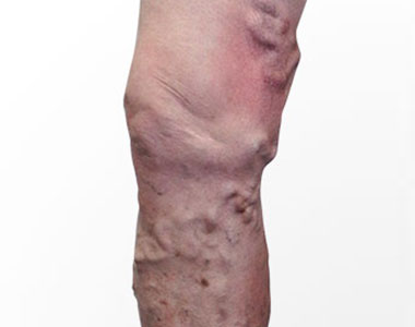
  • Persistent swelling of the leg or arm (edema)
  • Leg pain or tenderness while standing
  • Skin discoloration in the affected arm or leg
  • Veins in the arm or leg appearing larger than normal

Sometimes the development of a serious health complication such as pulmonary embolism is responsible for the symptoms in the DVT patients which are as follows:

  • Shortness of breath
  • Chest pain
  • Cough (blood may be coughed out as well)
  • Back pain
  • Profuse sweating
  • Lightheadedness
  • Blue nails or lips

These symptoms appear suddenly, and an immediate medical attention is required.

Diagnosis:

The diagnosis is initiated by a thorough clinical examination and involves review of the patient’s medical history. Based upon these findings, the physician will categorize the patient into low or high risk DVT. Further tests may be considered to rule out other health problems and to confirm the diagnosis, which are as follows:

  • Blood tests: These are considered to evaluate any blood disorders that increase risk of DVT.
  • D- dimer test: In this test, the levels of a protein called D-dimer is assessed. In patients with DVT, high levels of this protein can be found in the blood.
  • Duplex ultrasonography: It is a non-invasive evaluation of the blood flow in the arteries and the veins of a patient. But this test is not preferred for a thrombus located very deep inside the body such as pelvis.
  • Venography: It is a specialized type of an x-ray procedure wherein a dye is injected into a vein of the foot, and the blood clot if present is detected.
  • Magnetic resonance imaging (MRI): It is performed in patients for whom ultrasound examination is inappropriate or unfeasible. It can even detect a thrombus present in the patient’s pelvis and the thigh.

Prevention:

A recent World Thrombosis Day Ipsos survey showed that there is less awareness of DVT in countries around the world. Thus, the first step in prevention is making the public aware of DVT. The preventive measures are different depending on the person’s condition who are at risk of developing DVT:

During pregnancy

In pregnant women, certain medication may be prescribed if she has risk factors such as likelihood of having a cesarean birth, bed rest, history of DVT, and inherited thrombophilia.

During travel or in longer hours of sitting posture:

  • Drink lots of fluids.
  • Wear loose fitting clothes.
  • Walk and stretch at regular intervals.
  • Use special stockings to improve blood flow and to prevent blood clots but consult your health care provider before using it.

Treatment:

Treatment for DVT includes certain medicines and therapies, which are as follows:

Medications:

  • Anticoagulants: They prevent further formation of blood clots but don’t dissolve the already existing ones. Most common anticoagulants used are heparin and warfarin. Warfarin is not recommended during pregnancy.
  • Thrombin inhibitors: These are recommended for people who can’t take heparin.
  • Thrombolytic medicines: These are recommended only during life threatening condition.

Compression stockings:

It may be recommended to prevent swelling and to avoid post-thrombotic symptoms in the patients.

Surgical treatment:

Surgical treatment may be performed when medications fail to dissolve the clots.

Book Online Consultaion


    Deep Vein Thrombosis Blog

    DOWNLOAD PDF

    Subscribe the Hearty Life Blogs


      cci-chip_angioplasty.jpg

      Globally, coronary artery disease (CAD) is a common cause of death. Traditional coronary angioplasty bypass grafting (CABG) and percutaneous coronary intervention (PCI) are popular methods for managing CAD. PCI is a preferred intervention, while CABG is reserved for complex cases. However, some patients have associated risk factors that make them unsuitable for surgery.

      Cardiologists now use the CHIP (Complex, High-Risk Indicated) angioplasty approach for patients with complex, high-risk, and severe coronary disease..

      Who is eligible for CHIP angioplasty?

      Once after reviewing the medical condition of the patient, the CHIP program team will decide whether the person is a potential candidate for CHIP angioplasty. However, individuals who meet the following criteria are considered as potential candidates for CHIP angioplasty:

      • Advanced age- People with advanced age may not be able to tolerate bypass surgery and its complications.
      • History of kidney disease, stroke, or diabetes- The presence of co-morbid conditions may complicate the surgery and its outcome.
      • Location of CAD, including left main or bifurcated disease- The blood vessel is difficult to access and treat.
      • Chronic total occlusion (CTO) of the coronary arteries- CTO may cause sudden heart attack, and are dangerous.
      • History of open-heart surgery- Patients who have undergone open-heart surgery may be poor candidates for bypass surgery.
      • Advanced stage of heart failure- The failing may not be amenable to the stress of the bypass surgery.

      How is the treatment plan developed?

      The CHIP team will review and discuss the patient condition. Based on the extent of CAD (despite medical therapy), co-morbidities, and hemodynamic state, the treatment plan may include:

      • Surgical intervention
      • Conventional PCI
      • Protected PCI
      • Medical management

      The success of a CHIP program is dependent upon:

      • Highly skilled and experienced doctors
      • Advanced equipment
      • An expert and well-coordinated team of doctors, paramedics, nursing and support staff

      CHIP angioplasty techniques

      CHIP angioplasty comprises of advanced techniques like:

      • Rotational atherectomy: It is a common type of atherectomy device for plaque removal. Currently, this technique is used for ostial and heavily calcified lesions, which cannot be treated with balloon angioplasty.
      • Complex bifurcation stenting: This type of stenting is used to remove the blockage from the site where the blood vessel divides into two.
      • Specialized antegrade and retrograde chronic total occlusion (CTO) approaches.

      What are the benefits of CHIP angioplasty?

      CHIP angioplasty offers the following benefits:

      • Reduces symptoms
      • Improves the quality of life
      • Confers lower risk of re-hospitalization


      disease-condtions-thromboembolism.jpg

      Thromboembolism refers to a condition where a blood clot formed in the blood vessel unplugs itself and flows in the blood stream to block another blood vessel leading to obstruction of blood flow. Although clotting is a normal bodily function, if a clot occurs where it is not required, it may lead to serious problems.

      Types of Thromboembolism:

      The type of thromboembolism differs depending on the area in which the clot may appear. It can obstruct the blood flow in the veins, arteries, brain, gastrointestinal tract or kidneys.

      Thromboembolism can be further classified as below:

      Venous Thromboembolism:

      A venous thromboembolism is a thrombus (blood clot) formed in a vein. The blood flow in the veins is slow when compared to arteries which can increase the likelihood of the blood clots forming in these vessels.

      It can be categorized into three different types

      • Superficial Vein Thrombosis
        The clot may occur in a superficial vein, close to the surface of the body. Usually, it is not serious until it enters the deep veins.
      • Deep Vein Thrombosis (DVT)
        Deep vein thrombosis occurs within the deep veins, and mostly occurs within the legs.
      • Pulmonary embolism (PE)
        Pulmonary embolism is a medical emergency in which the thrombus blocks the blood vessel in the lungs.

      Renal Vein Thrombosis (RVT)

      A renal vein thrombosis is a thrombosis that occurs in the veins that drain blood away from the kidneys. These clots reduce the ability of the kidneys to clean and filter the blood.

      Arterial Thrombosis (atherothrombosis)

      Arterial thrombosis is much less common than venous thrombosis. It can have similar risks. Usually arterial thrombosis may lead to necrosis of the tissue.

      A thromboembolism in the coronary artery can cause a heart attack. If blood supply to the brain is disrupted, the patient may suffer a stroke.

      Symptoms:

      Signs and symptoms of venous thromboembolism include the following

      DVT

      • Pain in arm or leg, usually in the thigh or calf
      • Swollen leg or arm
      • Reddening or warming of the skin
      • Red streaks on the skin

      PE

      • Shortness of breath
      • Chest pain under your rib cage that can get worse when you take a deep breath
      • Rapid heart rate
      • Feeling lightheaded or passing out
      • Accentuated second heart sound

      Arterial Thrombosis

      • Muscle pain or spasm in the affected area
      • Numbness and tingling in the arm or leg
      • Pale color of the arm or leg (pallor)
      • Weakness of an arm or leg

      If the condition is severe, it may lead to blister formation and shedding of the skin, leading to tissue necrosis.

      Symptoms of a clot in an organ vary with the organ involved but may include:

      • Pain in the part of the body that is involved
      • Temporary decrease in organ function

      Causes:

      The blood clots can occur due to injury to a vein, consequence of a surgery, use of certain medications and lack of movement of the limbs.

      In the case of PE, the blood clot may block the blood vessels of the lungs. The affected portion of the lung may die due to loss of blood supply, the condition is called as pulmonary infraction, making it difficult to provide oxygen to the rest of the body.

      Occasionally, the blood vessels can be blocked by substances other than blood like collagen, part of a tumour, air bubbles etc.

      Risk factors:

      The common risk factors include

      • Inheriting a blood-clotting disorder
      • Prolonged immobility which may be due to bed rest or long trips
      • Injury or surgery
      • Pregnancy
      • Birth control pills or hormone replacement therapy
      • Being overweight or obese
      • Smoking
      • Cancer
      • Heart failure
      • Inflammatory bowel disease
      • Family history of deep vein thrombosis or pulmonary embolism

      Diagnosis:

      The diagnosis of thromboembolism includes the following tests which could be considered depending on the type:

      D-dimer: The blood sample is tested for the presence of D-dimer which is a marker for the presence of blood clots.

      Duplex Ultrasound: This is an imaging test in which the presence of clots is identified using ultrasound waves.

      Pulse oximetry: In this test, a sensor attached on the end of the finger of the patient helps to measure the level of oxygen in the blood.

      Arterial blood gas: The blood drawn from the artery is checked for oxygen levels present in it.

      Chest X-ray: This test may not be useful in finding the presence of clots, but can help to rule out a clot.

      Other diagnostic tests may include ELISA, pulmonary angiography, venography, echocardiography, helical computed tomography of pulmonary vessels etc.

      Treatment:

      Treatment of thromboembolism includes:

      Blood thinners: These are anticoagulant drugs which prevent formation of new clots while the body works to break up the previous clots present. They include heparin, low-molecular-weight heparin, apixaban, edoxaban, rivaroxaban, and warfarin.

      Clot-busting drugs: These are intravenous injections to dissolve the clots in the case of life-threatening situations. They include drugs belonging to the class of tissue plasminogen activators.

      Surgical and other procedures:

      • Removal of clot: Surgery is considered only if the condition is life threatening. The clot is removed using a catheter threaded through the blood vessel.
      • Vein filter: A catheter can also be used to position a filter in the inferior vena cava which helps to stop the clots from being carried into the lungs.

      Prevention:

      If the patient is at a risk of the condition, the following preventive measures may be considered to reduce the occurrence:

      • Use of blood thinners
      • Use of compression stockings
      • Use of intermittent pneumatic compression devices that automatically squeeze the legs to keep blood flowing

      Other preventive measures include

      • Regular exercise
      • Maintaining healthy weight
      • Cessation of smoking

      Consideration during travel include

      • Walk every 1 to 2 hours
      • Stretch legs and move around on the seat
      • Drink lots of fluids
      • Avoid smoking and consumption of alcohol before journey

      Book Online Consultaion


        Thromboembolism Blog

        DOWNLOAD PDF

        Subscribe the Hearty Life Blogs


          disease-condtions-varicose_veins.jpg

          Varicose veins are the twisted, swollen and bulging veins that appear in blue, red and skin color, just beneath the surface of the skin. They occur mostly in the legs and feet, causing aching pain and discomfort in the legs.

          Larger varicose veins appear rope-like and make the skin to bulge out. Inner varicose veins usually do not appear, but cause aching pain and swelling in the legs. Spider veins are the type of varicose veins which are most common and occur superficially, surrounded by the patches of flooded capillaries.

          Causes:

          Arteries have a thick layer of muscle or tissue to supply oxygenated blood to different parts of the body. Veins carry blood back to the heart and it should work against the gravity, so they rely on surrounding muscles pressure. Valves in the veins open to move the blood towards the heart and then close to prevent the back flow of blood.

          Any damage/abnormality in the functioning of the valves will lead to pooling up of blood in the vessels, causing them to stretch and twist, resulting in varicosity. It mostly occurs due to prolonged standing and walking, which builds up pressure in the veins of the lower legs.

          Signs and Symptoms:

          • Dark purple or blue color veins
          • Twisted and bulging veins that appear on the surface of the skin
          • Aching pain and heaviness in the legs
          • Burning, throbbing, muscle cramping and swelling in the lower legs
          • Worsened pain after sitting or standing for a long time
          • Itching around affected veins
          • Skin discoloration around a varicose vein
          • Web-like reddish blue varicose veins called spider veins

          Risk factors:

          The factors that increase the risk of developing varicose veins are:

          • Age: As the age increases, the functioning of the valves in the veins may be damaged, resulting in pooling up of blood in the lower leg veins, leading to varicose veins.
          • Gender: Hormonal changes in women during pregnancy, premenstruation and menopause will relax the vein walls, which increases the risk for varicose veins. Use of hormonal birth control pills also increases the risk.
          • Pregnancy: Blood volume increases during pregnancy to support the developing fetus, but can also produce a side effect of enlarged veins.
          • Family history: If any family member has varicose veins or deep vein thrombosis, it increases the risk.
          • Obesity: Being overweight will increase the pressure in the veins of lower legs.
          • Standing or sitting for long periods of time will reduce the free flow of blood in the blood vessels and results in clot formation.

          Complications:

          • Ulcers: Skin ulcers may form near the ankle, which is an indication of vascular disease.
          • Blood clots: Clots may occur in the veins deep inside the legs, causing swelling and leg pain. The condition is referred as thrombophlebitis.
          • Bleeding: Sometimes, the superficial veins may burst, causing bleeding.

          Diagnosis:

          A physical examination is performed, and the patient is enquired about the nature of leg pain diagnose varicose veins. The family medical history of varicose veins and information about their daily lifestyle activities is gathered. Certain tests may be needed to confirm the diagnosis of varicose veins, and include:

          • Ultrasound test: This is a non-invasive test, which uses sound waves to create the images of internal organs of the body. This test helps to identify the blood flow in the veins and also identify the clots and weakened or leaky valves in the veins.
          • Venogram: This test is a type of angiogram, which uses contrast dye to clearly view the blood flow through the veins, mainly if physician suspects the presence of large clots in the veins.

          Treatment:

          Initially, after the diagnosis of varicose veins, the doctor would recommend some lifestyle modifications that can relieve pain and reduce worsening of the condition, such as:

          • Perform exercises
          • Elevate the legs.
          • Avoid tight fitting clothes that can prevent the free circulation
          • Avoid long periods of standing or sitting

          Compression stockings: It will compress the leg veins and muscles and improve the blood flow efficiently, without forming any clots in the veins. The stockings are available in different sizes and lengths and can be bought without prescription in the local pharmacy.

          Sclerotherapy: This procedure is mainly used for severe varicose veins, and involves injecting a chemical solution near the varicose veins, which helps in closing and scarring of the veins.

          Laser treatment: This technique uses bursts of light reflected onto the vein, which makes the vein slowly fade and disappear. This test is mainly used to treat smaller varicose veins.

          Catheterization: This procedure uses a thin, flexible tube (catheter) that is inserted into the enlarged vein, which uses radiofrequency or laser energy, mainly to collapse the larger varicose veins.

          High ligation and vein stripping: Veins are tied off before it joins a deep vein and then it is removed through an incision.

          Ambulatory phlebectomy: Series of small punctures are made in the skin under general anaesthesia, to remove the smaller varicose veins.

          Endoscopic vein surgery: A thin tube attached with a camera is inserted into the vein to view and close the varicose veins and then removing them by making small incisions on the skin, under general anaesthesia.

          Prevention:

          Varicose veins cannot be prevented completely, but can be reduced by following some measures like:

          • Exercising regularly and losing extra weight
          • Eating a high-fiber and low-salt diet
          • Avoiding high heels and tight hosiery
          • Elevating the legs.
          • Changing the position regularly, when sitting or standing for long periods

          Book Online Consultaion


            Varicose Veins Blog

            DOWNLOAD PDF

            Subscribe the Hearty Life Blogs


              disease-condtions-pda.png

              Patent ductus arteriosus (PDA) is a congenital heart condition in which there is an opening between the pulmonary artery and the aorta. The ductus arteriosus is a small connection in the fetal heart helps oxygen rich blood to by-pass the immature baby’s lungs and flow into the body. This connection naturally closes shortly after birth. The connections that fail to close, are called patent ductus arteriosus, where the oxygen-rich and oxygen-poor combines together resulting in an increase in the workload of heart and various other complications.

              It is the sixth most common type of congenital heart disease, and is frequently diagnosed in infants; although it may remain unknown until childhood or even adulthood.

              Symptoms:

              The symptoms depend on the size of the patent duct and the gestational age of the neonate. A small patent ductus arteriosus can remain unrecognized until adulthood, whereas, a large patent ductus arteriosus may lead to life threatening conditions such as heart failure.

              In neonates, a heart murmur on examination with a stethoscope during regular checkup may indicate PDA.

              In infants, a large PDA would show the following symptoms:

              • Inability or difficulty in feeding, leading to poor growth
              • Sweating on crying or eating
              • Persistent fast breathing or breathlessness
              • Cough
              • Lower respiratory tract infections
              • Pneumonia
              • Easy tiring
              • Rapid heart rate

              The symptoms in the case of adults with undiagnosed PDA include heart failure, fast heart beating (atrial fibrillation) or with the reversal of shunting – unoxygenated blood from the right side of heart to the left side of heart.

              Causes:

              In most of the children the cause of PDA is unknown, but genetic factors are thought to play a causative role. 

              Every baby is born with ductus arteriosus which eventually narrows and closes within three to four days after birth. It may take longer time for closure in the case of premature babies. 

              But if the duct doesn’t close, it may lead to an increase in the blood flow to the heart and lungs of the baby, which might enlarge or weaken the heart muscle.

              Risk factors:

              The risk factors of having patent ductus arteriosus include:

              • Gender: PDA is more common in girls than in boys.
              • Prematurity: It is more common in babies who are born before the gestational term than in full term babies.
              • Family history: A family history of heart conditions and other genetic disorders, such as Down’s syndrome may increase the risk of PDA.
              • Rubella infection: If the mother is affected with rubella infection during pregnancy, the virus may cross the placenta and spread through the baby’s circulatory system potentially damaging blood vessels and organs including the heart.
              • High altitude birth risk: Babies born at altitudes higher than 10,000 feet are at an increased risk.
              • Congenital heart problems: Babies with congenital heart problems such as hypoplastic left heart syndrome, transposition of the great vessels, and pulmonary stenosis are at high risk.

              Diagnosis:

              A heart murmur heard during a regular checkup may lead to further work-up for PDA.

              The following tests Are recommended for diagnosis of PDA:

              • Chest X-ray to assess the condition of the heart and lungs, and also to rule out other conditions
              • Echocardiogram to assess the heart, its valves and chambers for any defects and see if the heart is pumping properly
              • Electrocardiogram (ECG) to assess the electrical activity of the heart to diagnose any heart defects or rhythm problems
              • Cardiac catheterization to rule out other congenital heart defects found during an echocardiogram or in a case where the catheter procedure is considered for the treatment.

              Treatment:

              Treatment is generally not considered, as the PDA usually closes on its own in the case of a premature baby. Close monitoring is considered for full term babies, children and adults with small PDA and with no other health complications. During follow-up, if the baby does not have any other complications, it is considered to be closed. On the other hand, if the baby has certain heart problems or defects, the ductus arteriosus might be lifesaving.

              In a premature baby, it may take one to two years. But in full term babies, PDA that remains open after several weeks rarely closes on its own.

              Medicines such as indomethacin or ibuprofen may be considered. These medicines work well for some newborns. The earlier treatment is given the greater are the chances for it to succeed.

              Medical procedures may be considered in the case where medications are not effective. 

              A transcatheter procedure is used to block blood flow through the vessel. This is usually using an umbrella device that closes the PDA. This procedure does not require general anesthesia and requires hospital stay less than 24 hours. 

               An open heart surgery might be considered if the catheter procedure is not suitable to repair PDA. This is usually reserved for very large ductus and other associated heart defects.

              Prevention:

              Patent ductus arteriosus may not be prevented, but certain measures should be observed for a healthy pregnancy.

              • Early prenatal care
              • Healthy diet
              • Regular exercise
              • Avoiding consumption of alcohol, illegal drugs and cigarettes.
              • Getting vaccinated as recommended.
              • Optimal diabetes control.
              English Video

              Book Online Consultaion


                Patent Ductus Arteriosus Blog

                DOWNLOAD PDF

                Subscribe the Hearty Life Blogs


                  disease-condtions-percutaneous_coronary_intervention.jpg

                  • Coronary arteries are the vessels which supply blood (oxygen and nutrients) to the heart. With age, cholesterol and fats (called plaques) are deposited on the inner walls of the arteries (atherosclerosis), thereby narrowing the lumen of the arteries.
                  • The deposition of plaque is more likely at the site where the main coronary artery branches (bifurcation) due to the forces associated with the changes in blood flow. Narrowing (or stenoses) of the main coronary artery and the adjoining side-branch vessel is called bifurcation lesion or bifurcation blockage.
                  • Treating bifurcation lesions is more challenging than treating the blockage of the blood vessels that do not involve branches, as there are no stents with “Y” configuration available. Percutaneous coronary intervention is the current treatment option recommended for bifurcation lesions.
                  • Percutaneous coronary intervention (PCI) is a minimally invasive, non-surgical procedure that involves placing a stent (a small metal mesh) using a catheter (a thin, flexible tube) to treat the blockages in the arteries. For bifurcation blockages, two stents may be placed simultaneously.

                  What are the complications of PCI procedure for bifurcation lesions?

                  The complications associated with PCI procedures for bifurcation lesions include:

                  • Low success rate
                  • Complete obstruction of the artery caused by the formation of thrombus in the first month after implantation (subacute stent thrombosis)
                  • Recurrence of the blockage or the narrowing of the blood vessels (restenosis)
                  • Periprocedural myocardial infarction
                  • Stent deformation
                  • A gap between the stent and the arterial wall, which is greater than the stent thickness (mal-apposition)

                  What are the different types of PCI techniques performed for bifurcation lesions?

                  The following are the different types of PCI techniques used for bifurcate lesions. These techniques may vary in different cases.

                  T stenting technique: This technique involves positioning two stents in a ‘T’ shape. In this technique, the first stent is placed in the side branch close to the ostium (opening of the side branch), while the main branch is inflated with a balloon at low pressure. Then the second stent is placed in the main vessel.

                  Reverse T-stenting: This is a modified method of the classical T stenting technique. In this method, the first stent is placed in the main vessel crossing the side branch, and the second stent is placed in the side branch.

                  *Note: T-stenting and reverse T-stenting techniques are considered when the angle between the side branch and the main vessel is >70 degrees.

                  Culotte technique: In this method, the first stent is deployed into the vessel with the sharpest angulation, which covers both the main vessel and the side branch. Then, a second stent is passed through the struts of the first stent and is positioned in the main vessel only. This technique allows complete coverage of the lesion and has low rates of in-stent restenosis and periprocedural myocardial infarction.

                  Crush technique: In this technique, the two stents are passed at the same time in both the vessels; the stent in the main vessel more proximal than the stent in the side branch. First, the side-branch stent is paced, ad its wire and balloon are removed. Then, the main vessel stent is positioned, which flattens the protruding part of the side branch stent.

                  Simultaneous kissing technique: In this method, the two stents are advanced into the side branch and the main vessel and the stents are simultaneously positioned. This positioning of the two stents creates a new carina (the inflection point where the side branch separates from the main branch) in the proximal portion of the main vessel. This technique is considered only when the proximal vessel can accommodate both stents.


                  disease-condtions-device_closure_for_vsd.jpg

                  The heart consists of four chambers, of which upper two chambers are called as atria and lower two chambers are called as ventricles. Right and left chambers are separated by a wall of muscle called a septum. The septum separating the atria is called inter atrial septum and the once between the two ventricles in inter ventricular septum.

                  Right two chambers – right atrium and right ventricle pump the deoxygenated blood collected from various parts of the body to the lungs. Blood gets saturated with oxygen in the lungs and returns back to the left side of the heart.

                  Ventricular septal defects (VSD) is a common type of congenital heart defect, which is characterized by an abnormal opening or a hole in the interventricular septum, the dividing wall between right and left ventricles. The oxygen-rich blood from the left ventricle will enter into the right ventricle through the opening, thereby getting mixed with deoxygenated blood which then enters into lungs. This will force the heart and lungs to work harder.

                  Causes:

                  The exact cause of VSD is unknown. During fetal heart developmental stage, the heart develops from a large tube which eventually divides into chambers and walls. Any abnormality in this process will lead to the formation of a defect in the septum. If the defect is in the interventricular septum, then it is said to be ventricular septal defect. There may be one or more VSDs.

                  ventricular septal defects

                  Types:

                  Based on the location and development of VSD, it is classified into following types:

                  • Conoventricular Ventricular Septal Defect: It occurs just below the pulmonary and aortic valves.
                  • Perimembranous Ventricular Septal Defect: It occurs in the upper part of the ventricular septum.
                  • Inlet Ventricular Septal Defect: It occurs adjacent to the tricuspid and mitral valves. This type of defect might be associated with atrial septal defect.
                  • Muscular Ventricular Septal Defect: It is the most common type of VSD, which occurs in the lower muscular part of the interventricular septum.

                  Signs and symptoms:

                  Small defects in the septum do not show any symptoms because it closes on its own gradually during childhood.

                  Large defects show signs and symptoms usually after birth within a few weeks or months.

                  The first sign of VSD is heart murmur, which is a whooshing sound that can be heard using a stethoscope. The other symptoms include:

                  • Fatigue (tiredness)
                  • Arrhythmias (abnormal heart rhythm)
                  • Fast breathing or breathlessness
                  • Poor feeding
                  • Poor weight gain
                  • Pale skin
                  • Enlarged liver

                  Risk factors:

                  Ventricular septal defects mostly occur due to defective genes and chromosomes, that may be hereditary.

                  Environmental factors during pregnancy may also play a role in development of VSD in the fetus.

                  Complications:

                  Large or medium septal defects if left untreated, may lead to life threatening complications such as:

                  • Heart failure, as heart need to work harder to pump enough blood to the body.
                  • Pulmonary hypertension (increased blood flow to the lungs results in increased blood pressure in the lung arteries).
                  • Endocarditis (infection in the endocardium of heart).
                  • Other heart problems such as abnormal heart rhythms and valve problems.

                  ventricular septal defects

                  Diagnosis:

                  If heart murmurs are detected during the physical examination, the patient may be advised further testing to conform the diagnosis.

                  • Echocardiogram: This test is the primary tool for the diagnosis of VSD, as it can be used to determine the size, location and severity of the ventricular septal defect. Sound waves are used to produce the detailed images of the heart.
                  • Electrocardiogram (ECG): This test records the electrical activity of the heart and helps to identify the abnormal heart rhythms and defects in the septum.
                  • Chest X-ray: X-rays are used to view the images of heart and lungs. In VSD, this test helps to determine the enlarged heart and extra fluid in the lungs.
                  • Cardiac catheterization: The test involves inserting a thin, flexible tube into the blood vessel at the groin or an arm, which is guided to the heart, to identify any congenital heart defects and to determine the function of the heart chambers and its valves.
                  • Pulse oximetry: Oxygen levels in the blood can be measured using a small clip which is placed on the fingertip.

                  Treatment:

                  Usually treatment is not needed for small VSDs, as they close on their own gradually after birth. 

                  Babies with larger VSD need surgery to prevent any further complications. 

                  The treatment for ventricular septal defects include:

                  • Medications such as diuretics like furosemide are used to reduce the amount of fluid in the blood that is pumped to the lungs and in the circulation. Beta blockers like metoprolol, propranolol and digoxin are used to maintain the regular heartbeat.
                  • Surgical procedures include surgical repair, catheter procedure and hybrid procedure.
                    Surgical repair involves open heart surgery, in which the doctor uses a patch or stitches to close the hole, performed under general anaesthesia.
                    In catheter procedure, a catheter is inserted into a blood vessel and then passed to the heart. The defect is closed using a specially sized mesh device.
                    Hybrid procedure uses both surgical repair and catheterization procedures to close the hole, with the help of a heart-lung machine and a catheter placed through an incision.

                  Prevention:

                  Ventricular septal defects cannot be prevented, but following certain measures during pregnancy may be helpful to prevent the risk of VSD. The measures include:

                  • Prenatal care before pregnancy: Consult a physician before planning for pregnancy and inform him about the family history of any congenital defects and the medications using currently, so that he may give suggestions or recommend some lifestyle modifications to avoid the risk of heart defects.
                  • Healthy diet: Having a balanced diet including vitamins and folic acid during pregnancy will help in giving birth to a healthy child without any heart defects.
                  • Regular exercise under the supervision of a gynecologist is necessary during pregnancy.
                  • Avoid alcohol, tobacco and harmful drug use : during pregnancy to prevent the risk of VSD.
                  • Get vaccinated: Check whether you are vaccinated for vaccine-preventable infections before getting pregnant.
                  • Control diabetes: Monitor your sugar levels regularly to prevent the risk of heart defects.
                  English Video

                  Book Online Consultaion


                    Ventricular Septal Defects Blog

                    DOWNLOAD PDF NOW

                    Subscribe the Hearty Life Blogs


                      disease-condtions-device_closure_for_asd.jpg

                      What is an Atrial Septal Defect or ASD? 

                      Heart consists of four chambers, of which upper two chambers are called as atria and lower two chambers are called as ventricles. Atrial septal defect (ASD) is a type of congenital heart defect, in which there is an abnormal opening or a hole in interatrial septum (dividing wall between two atria). This opening allows the passage of pulmonary venous blood from left atrium to right atrium, causing mixing of oxygenated and deoxygenated blood in right atrium and increasing the flow of blood to lungs. The increased blood flow may increase the workload of the lungs, and eventually cause damage to heart and lungs.

                      What causes an ASD?

                      The exact cause of ASD remains unclear. However, it is believed that during fetal developmental stages, a hole is present in the interatrial septum, which gradually closes before birth or during infancy. If the hole persists, it is called an atrial septal defect.

                      Types of Atrial Septal Defect or ASD?

                      Based on the location and development of ASD, it is classified into four types:

                      • Ostium secundum ASD: It occurs in the middle part of the interatrial septum. This is the most common type of ASD and accounts for 75% of all atrial septal defects. This type of ASD is commonly detected in adults in their third and fourth decades of life. Some can be detected in children when an abnormal heart sound is detected at the time of a routine health check or vaccination visit. 
                      • Ostium primum ASD (20%): It occurs in the lower part of the interatrial septum, adjacent to atrioventricular (AV) valves. It usually occurs as a part of other congenital heart defects. This defect is usually detected in early life as this is associated with many complications.
                      • Sinus venosis ASD (4%): It occurs in the upper part of the interatrial septum, near the veins that drain into the right and left atrium. This is usually identified in third and fourth decade adults. 
                      • Coronary sinus ASD (<1%): It occurs in the interatrial septum between the coronary sinus and the left atrium. This is very uncommon and patients are asymptomatic.

                      Signs and symptoms

                      Usually after birth, babies who have ASD may not show any associated signs and symptoms. But, symptoms may appear during adulthood around the age of 30 years. Most of them don’t have any symptoms even after many years.

                      Some of the common symptoms associated with ASD are:

                      • Heart murmur, a swishing sound that can be heard through a stethoscope
                      • Heart palpitations
                      • Arrhythmias (abnormal heart rhythms)
                      • Fatigue (feeling tired mainly after exercise)
                      • Shortness of breath
                      • Swelling of legs, feet or abdomen
                      • Stroke
                      • Lung infections such as pneumonia

                      Risk factors:

                      Genetics and environmental factors usually play a role in congenital heart defects. Some conditions during pregnancy may confer high risk for heart defects, 

                      Complications of an untreated ASD:

                      • Right-sided heart failure
                      • Arrhythmias
                      • Increased risk of a stroke
                      • Shortened life span
                      • Pulmonary hypertension (increased blood flow to the lungs results in increased blood pressure in the lung arteries)
                      • Eisenmenger syndrome (pulmonary hypertension causing permanent lung damage)

                      Diagnosis:

                      Most ASDs are diagnosed incidentally during regular heath check-ups. If a heart murmur is heard during auscultation, you may be advised to undergo further tests to confirm the diagnosis of ASD: 

                      • Echocardiogram: It is a specific diagnostic test for ASD, and provides a detailed image of the heart and blood flow through its chambers. The echocardiogram may show the blood flow through the interatrial septum and the size of defect in the septum.
                      • Chest X-ray: It helps to identify enlarged heart and lung changes.
                      • Electrocardiogram (ECG): It involves recording the electrical activity of the heart, to help identify arrhythmias.
                      • Cardiac catheterization: A thin, flexible tube called catheter is inserted into the blood vessel at the groin or arm and is guided to the heart. This test is helpful to determine the function of heart and its valves, and to measure the blood pressure in the lungs.
                      • Magnetic resonance imaging (MRI): It uses magnetic and radio waves to create three dimensional images of the heart and other organs. This test is recommended if the ASD is not clearly diagnosed with echocardiogram.
                      • Computerized tomography (CT) scan: It uses a series of X-rays to create detailed images of heart; mainly used if echocardiogram doesn’t help to diagnose ASD clearly.

                      Treatment:

                      Treatment of ASD depends on the age at diagnosis and the size, location and severity of the defect. Small ASDs might not need any treatment because it closes on its own. The doctor may recommend surgery if the atrial septal defect is large, even with fewer symptoms to prevent problems later in life. Treatment of ASD includes medications and surgery.

                      • Medications usually do not help in closing the hole, but reduces the signs and symptoms associated with ASD and risks of complications after surgery. Drugs such as beta blockers (to maintain regular heartbeat) and anticoagulants (to prevent the formation of blood clots) are used.
                      • Surgery includes cardiac catheterization and open-heart surgery.

                      Device closure of ASD

                      In cardiac catheterization, the doctor closes the hole in the septum with a mesh patch or a plug in the defect place using a catheter. The heart tissue then grows around the mesh slowly, sealing the hole permanently. This procedure is mainly performed to repair only the secundum type of atrial septal defects. Defects not amenable to closure with device may need open-heart surgery.

                      In open-heart surgery, the defect may be closed with stitches or a special patch. This procedure is done under general anesthesia, mainly for repairing primum, sinus venosus and coronary sinus atrial septal defects.

                      Prevention of ASD:

                      Atrial septal defects cannot be prevented, but following certain measures during pregnancy might be beneficial to prevent the risk of ASD, such as:

                      • Immunity test for rubella: If the person is not immune to rubella, it is necessary to get vaccinated.
                      • Monitoring current health conditions and medications use: Pre-existing health conditions and usage of any medications should be carefully monitored during pregnancy to prevent the risk of ASD.
                      • Reviewing family medical history: If a person has a family history of congenital defects, it is advised to visit a genetic counsellor before becoming pregnant to know the risks of atrial septal defects.
                      English Video


                      Our Specialities


                      • 24 hours emergency services
                      24 hours emergency services
                      • Clinics- weekly basis/monthly basis/ Yearly basis
                      Clinics- weekly basis/monthly basis/ Yearly basis
                      • Prevention of cardiovascular diseases
                      Prevention of cardiovascular diseases
                      • Diagnosis
                      Diagnosis

                      BOOK AN APPOINTMENT

                        dr. raghu

                        DR. RAGHU

                        MD; DM; FESC; FACC; FSCAI

                        Cardiology Coronary, Vascular and

                        Structural Interventions


                        Conditions & Diseases

                        Coronary angioplasty

                        Angioplasty

                        Coronary angioplasty

                        Aortic Stenosis

                        Coronary angioplasty

                        Atrial Fibrillation

                        Coronary angioplasty

                        Atrial Septal Defect


                        View More Services


                        disease-condtions-coronary_artery_disease.jpg


                        Coronary artery disease (CAD) is the most common type of heart disease, in which the coronary arteries ( blood vessels that supplies oxygen and nutrients to the heart muscle) become rigid and narrowed due to buildup of plaque (deposition of cholesterol) in the inner walls of the heart. This restricts the flow of oxygen rich blood to the heart muscle, which may result in angina (chest pain) and shortness of breath. If the plaque ruptures or due to a complete blockage in the artery, it may result in heart attack or sudden cardiac death.

                        Causes:

                        Build of cholesterol plaques within the blood vessels is termed atherosclerosis.

                        The plaque makes the inner walls of the artery sticky, so that the inflammatory cells, lipoproteins, and calcium travelling through the blood stream and adhere to the plaque. The plaque gradually enlarges, narrowing the blood vessel lumen, restricting blood flow to the heart muscle and thus reducing the supply of oxygen and nutrients to the heart.

                        Signs and symptoms:

                        • Angina (chest pain) – on exertion and in severe cases even at rest
                        • Shortness of breath – on exertion and in severe cases even at rest
                        • Palpitations (irregular heartbeats or skipped beats)
                        • Tachycardia (fast heartbeat)
                        • Weakness or dizziness
                        • Nausea
                        • Sweating
                        • Fatigue, mainly after exercise or any activity

                        Risk factors:

                        • Age: Risk for damaged or narrowed arteries increases with age.
                        • Gender: Men are at greater risk of developing CAD. In women, risk increases after menopause.
                        • Family history: Risk increases if any family member has coronary artery disease.
                        • Smoking: People who have the habit of smoking and who are exposed to smoke are at increased risk of CAD.
                        • High blood pressure: It results in the narrowing and hardening of the walls of arteries.
                        • High blood cholesterol levels: It can increase the risk of atherosclerosis.
                        • Diabetes: High degree of association – 2 to 4-fold greater risk
                        • Obesity: Overweight increases the risk of CAD.
                        • Life style activities: Reduced physical activity, high stress and unhealthy diet also contribute to the development of CAD
                        • Mental Stress: Responsible for young heart attacks Other possible risk factors, according to research, include:
                        • Sleep apnea: It is a condition in which the person may experience intermittent cessation of breathing during sleep, resulting in reduced oxygen levels in the blood. Sleep apnea may cause an increase in the blood pressure and high strain on the heart, leading to CAD.
                        • Increased C-reactive protein (CRP) levels in blood: CRP levels are increased mostly if there is any inflammation in the body. It is a risk factor for CAD.
                        • High triglycerides (a type of lipid).
                        • High homocysteine (amino acid) levels.
                        • Preeclampsia (high blood pressure during pregnancy).
                        • Alcohol use can lead to heart muscle damage.
                        • Autoimmune diseases such as Rheumatoid arthritis and Lupus.

                        Complications:

                        If left untreated, CAD can lead to life threatening complications such as:

                        • Angina:  Due to narrowed blood vessels, the heart muscles receive inadequate oxygen laden blood, causing chest pain or shortness of breath.
                        • Heart attack: The plaque may rupture forming a clot and blocking the blood flow to the heart muscle.
                        • Heart failure: Due to reduced oxygen and nutrient rich blood flow, the heart is unable to pump enough blood, thus increasing its workload. Over time, it may lead to weakening of heart muscle, also called heart failure.
                        • Arrhythmia: Reduced amount of blood supply may lead to abnormal heart rhythms
                        • Sudden cardiac death

                        Diagnosis:

                        The diagnostic work up includes a physical examination, in addition to medical history, family history, risk factors and any signs and symptoms of CAD. Further diagnostic tests may be recommended:

                        • Electrocardiogram (ECG): It records the heart’s electrical activity and helps to determine the blood flow in the heart as well as heart muscle stress.
                        • Echocardiogram: It uses sound waves to produce clear images of the heart and helps to identify the damaged heart muscles and valves.
                        • Stress test: The test involves performing any high intensity activity like walking fast on a treadmill or bicycling under medical supervision, and close monitoring of heart function during the physical activity. Symptoms, ECG, blood pressure and heart rhythm are closely monitored during the test. Based on the findings the probability of having significant coronary artery disease would be determined. This test has many limitations and fallacies.
                        • Cardiac catheterization and angiogram: A special medication called contrast is injected into the coronary blood vessels using a thin, flexible tube called catheter. The catheter is inserted through a small incision at the groin or through the wrist to visualise the blood vessels supplying the heart.
                        • Heart scan: Computerized tomography (CT) technologies are used to identify the calcium deposits in the blood vessel of the heart, which indicates CAD.

                        Coronary Artery Disease Treatment:

                        Lifestyle changes:

                        • Quit smoking
                        • Avoid processed foods and eat high protein and fiber rich diet
                        • Exercise regularly and lose excess weight
                        • Manage stress
                        • Control diabetes and blood pressure

                        Medications:

                        The doctor may prescribe medications, which help in reducing symptoms and severity of disease such as:

                        • Dyslipidemic agents such as statins, ezeimibe to lower the blood cholesterol levels
                        • Aspirin, a blood thinner, to prevent blood clots’ formation that might obstruct the coronary arteries
                        • Beta blockers decrease the heart rate and blood pressure and reduce the oxygen demands of the heart.
                        • Calcium channel blockers to lower blood pressure and improve blood supply to the heart.
                        • Trimetazidine and Ranolazine are anti-anginal drugs to reduce chest pain
                        • Nitroglycerin to dilate the coronary arteries and reduce chest pain
                        • Angiotensin-converting enzyme (ACE) inhibitors and angiotensin II receptor blockers (ARBs) to decrease blood pressure and improve heart efficiency.

                        Surgical procedures:

                        • Angioplasty and stent placement: It is also called as percutaneous coronary revascularization, and involves inserting a thin, flexible tube (catheter) into the narrowed coronary artery, followed by a wire with deflated balloon to the narrowed area. The balloon is inflated to widen the blood vessel; a stent may also be placed to keep the artery patent.
                        • Coronary artery bypass surgery: A graft is created using any blood vessel in the body, to bypass the blocked coronary arteries.

                        Prevention:

                        To maintain healthy arteries and to prevent coronary artery disease, some measures need to be taken such as lifestyle modifications which includes avoiding smoking and alcohol use, exercising regularly to maintain healthy weight, controlling sugar and cholesterol levels in blood and also eating a healthy diet.

                        Book Online Consultaion


                          Coronary Artery Disease Blog

                          DOWNLOAD PDF

                          Subscribe the Hearty Life Blogs


                            disease-condtions-acute_myocardial.jpg

                            • Acute myocardial infarction (AMI) is the necrosis of the heart muscles
                              resulting from acute obstruction of a coronary artery.
                              “Time is Muscle” in AMI .
                            • Primary angioplasty is a minimally invasive procedure that uses treats the
                              obstructed areas of the coronary arteries causing a myocardial infarction
                            • Considering long travel distances and lack of cath lab in many districts –
                              “Lyse now and stent soon” is the apt strategy for managing AMI.

                            Approach for managing AMI:

                            Benefits of Primary angioplasty:

                            • Class I therapy for AMI
                            • Preserves heart muscles
                            • Immediate restoration of blood flow
                            • Fast recovery
                            • Saves lives, immediate and long-term benefits

                            Procedure of primary angioplasty:

                            1. A balloon catheter is inserted into the main artery in the groin or arm
                            2. The catheter is passed gently into the aorta
                            3. The balloon is inflated at the narrowed area(s) of the artery, thereby opening and widening it.
                            4. A stent may also be placed to keep the artery open


                            +91 95424 75650

                            Call us now if you are in a medical emergency need, we will reply swiftly and provide you with a medical aid.


                            +91 95424 75650


                            Call us now if you are in a medical emergency need, we will reply swiftly and provide you with a medical aid.


                            Dr. Raghu | Heart Specialist in Hyderabad
                            Yashoda Hospitals, Sardar Patel Rd, behind Hari Hara Kala Bhavan, Kummari Guda, Shivaji Nagar, Secunderabad, Telangana 500003


                            Copyright © 2023, Dr. Raghu. All rights reserved.

                            [gtranslate]