Acute myocardial infraction is a sudden obstruction in the blood flow to the heart muscle.
Acute myocardial infraction is a sudden obstruction in the blood flow to the heart muscle.
Acute myocardial infraction is a sudden obstruction in the blood flow to the heart muscle. It is an emergency condition in which the plaque formed due to the deposition of fat, cholesterol, calcium, or cellular waste products is ruptured leading to the formation of thrombus. This leads to the obstruction of the coronary vessels resulting in an acute reduction of blood supply.

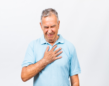
The symptoms of Acute myocardial infraction include:
The important signs of the patient to be considered include the following:
Coronary artery blockage is the primary cause of acute myocardial infarction. It is due to the accumulation of cholesterol in the arteries. The increase in LDL levels (bad cholesterol) in the body leads to plaque formation in the arteries. Saturated and trans fats are other types of fats which also lead to the formation of plaque in the arteries and obstruct the blood flow. Certain dairy products including butter and cheese, meat, beef, and processed foods are the main sources of saturated and trans fats.
During a heart attack, the plaque gets ruptured and spills cholesterol into the bloodstream. This leads to the formation of blood clots, when are big enough can block the artery at the site of rupture and deprives the heart muscle from obtaining enough oxygen and nutrients. There can be a partial block of the artery or a complete block of the artery. A complete block refers to an ST level elevation Myocardial Infraction (STEMI) and a partial block refers to a non-ST elevation Myocardial Infraction (NSTEMI).
Some of underlying risk factors of the acute myocardial infraction are modifiable.
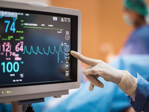
Electrocardiogram
Electrocardiogram remains a crucial tool used to diagnose a patient with acute myocardial infraction. One of the significant finding is the presence of the raised ST segment.
Cardiac Imaging
The coronary cardiac imaging is used to check for the presence or to rule out the coronary artery disease. The test is considered for the individuals who are at risk of having an acute myocardial infraction.
The laboratory tests should include identification of marker known as cardiac troponins, complete blood picture, lipid profile, renal function and metabolic panel.
Any patient diagnosed with acute myocardial infraction advised to take aspirin 165 mg – 325 mg immediately regardless of their condition (STEMI or NSTEMI). In STEMI the patient should receive dual antiplatelet agents, including heparin infusion. This should immediately be followed by a reperfusion with the percutaneous coronary intervention (PCI). If PCI is unavailable within 90 minutes of diagnosis of STEMI, an intravenous thrombolytic agent should be considered for reperfusion. NSTEMI in a stable asymptomatic patient can be managed with antiplatelet agents.
Any individual can improve heart health and prevent the occurrence of current heart condition by following the below-mentioned lifestyle changes:
Your heart needs to work 24/7 to keep your body systems working. Like any other body tissue, the heart too needs oxygen and nutrients to function efficiently. For this, we have a network of arteries that supply blood to the heart muscles which are called the coronary arteries. There are two chief coronary arteries:

the left and right coronary arteries, that branch out from the aorta near the point where the aorta and the left ventricle meet.
The right coronary artery supplies blood to the right atrium, the right ventricle, a small bottom area of the left ventricle and the back portion of the septum.
The left coronary artery supplies blood to the left atrium and ventricles and the front portion of the septum.
These coronary arteries give out various branches that supply blood to different parts of the heart.
When there is high level of unhealthy fats in the blood, they gradually start depositing in the insides of the coronary artery vessel wall and form a fatty plaque(atherosclerosis). This gradually narrows the lumen of the coronary arteries which obstructs blood supply to the heart. This condition is called as coronary artery disease. disruption of blood supply to the heart gives rise to a cluster of symptoms, the most important being chest pain or angina.
There are an array of heart diseases but they do share some common symptoms. If you experience any of these symptoms, it is perhaps a good idea to get yourself tested. Check these out here:

For continuous circulation, the left and the right side of the heart must work together. Here are the series of steps that causes the blood to flow in the heart, lungs and body.

Life is precious and to live life to the fullest, one should pay a little attention to his/her health- physical and mental. So, what do we do to be healthy- eat right and exercise, isn’t it? Actually, that isn’t it, though diet and exercise are very important for fitness but there is many more measures that we should take to keep ourselves healthy in and out. So, let us understand them one by one:
Ensuring that what goes inside your body is right- is the best thing you could do for your body. For a healthy heart, a 2000 calorie diet is recommended. Keep these points in mind:

All of us should spare atleast 30 minutes each day for exercise. Exercise doesn’t necessarily mean you should hit the gym, it means you need to keep your body moving- do brisk walk in the park, do yoga, or the best option would be, switch on your favorite music and groove on the beats.

In addition to exercises, inculcating physical activity in your daily routine like using stairs instead of the elevator, parking your car further and walking down to office, using a manual bike to go to office, or having standing desks to work on.

Obesity is a killer. You need to keep your weight in check. The best way to find out if your weight is just right is to measure the body mass index.The BMI calculation is very easy, divide your weight in kilograms by their height in metres squared. If your BMI is:

Go for regular health checks. Remember these numbers:

Smoking is one of the chief risk factors for not only heart disease but also deadly cancers, lung diseases and chronic disorders like high blood pressure. Quitting smoking is one of the best gift that you could give your body today.

People with stress and mental illnesses like depression are at greater risk of heart diseases. Have a good social life, spend time with friends and family, explore new places, give some time to your hobbies and interests- do anything that relaxes you. A relaxed mind and body makes you a more productive person and helps you stay healthy.

Research suggests laughing lowers stress hormones, decreases inflammation in your arteries, and raises good cholesterol levels.

Heart is a complex organ and can get affected by diseases that can affect various systems of the heart. The common heart ailments that would be observed include:
Coronary Artery Disease (CAD) – accumulation of cholesterol plaques within the walls of the blood vessels (coronary arteries) supplying the heart. This leads to obstruction to blood flow of the heart that can cause chest pain or heart attack.
Valvular heart disease: Heart valves are flap-like structures akin to doors between rooms. They control the blood flow between various chambers of the heart. They play a key role in blood circulation.
There are four valves in the heart
These valves can either get narrowed (stenosis) or get “leaky” (regurgitation).
Cardiomyopathy: This disease affects the heart muscle leading to inefficient heart pumping efficiency. Cardiomyopathy can be either due to:
Doctors usually recommend a battery of tests based on the system of the heart that has been affected. Based on a detailed history followed by a detailed physical examination, appropriate battery of tests would be suggested. The diagnosis of the heart ailment is critically dependent on the results of tests.
In addition to confirming the diagnosis, test results might indicate the disease complications and thus your doctor is able to stage the disease and the possible outcomes.
This test detects and records the electrical activity of the heart. This is a simple, non-invasive test which is very useful to determine abnormalities in the heart rate, rhythm and to identify risk of damaged heart muscle or other structural changes in the heart. This test detects the presence of arrhythmias and coronary artery disease.
Exercise makes your heart work harder. Exercise stress testing is done either on a treadmill or cycle ergometry with the patient connected to an electrocardiogram. Exercise stress testing may identify myocardial ischaemia, haemodynamic/ electrical instability, or other exertion-related signs or symptoms. When an individual is not able to exercise, medications are given to stress the heart and the response is evaluated.
Chest X ray is very useful to differentiate whether shortness of breath is due to a respiratory disease or heart disease. It can also help in detecting complications of heart failure such as cardiomegaly, interstitial oedema, pulmonary oedema and pleural effusions.
Coronary angiography is useful to determine the health of the coronary arteries. In this test, a catheter is inserted into the coronary arteries and a dye is injected to produce clear X ray images of the coronary arteries. This helps to find out the presence, location and extent of vessel narrowing. The results also help to decide which type of treatment would be most appropriate for correction of heart problem.
This test gives an ultrasound image of the heart. Echocardiography can provide information about the size and shape of heart chambers, blood flow velocities, heart muscle function when they contract and relax, abnormalities of the movement of the heart wall, valve function, and presence of thrombus (blood clot) in the heart.
Stress echocardiography helps in detecting decreased blood flow to heart during exertion. In this test, echocardiography is done immediately post stress. The stress can be exercise or could be induced by medications.
MPS is a non-invasive test which helps to determine how well blood flows through your heart muscles. In this test, a small amount of a radioactive substance is injected into the blood. The test evaluates the severity of coronary artery disease and provides guidance regarding the need as well as success of invasive procedures like angioplasty and stent insertion.
Cardiac CT provides detailed images of the heart. This helps to identify structural abnormalities in the heart and blood vessels such as aneurysms, valve dysfunction and damage to the pulmonary vasculature. Cardiac CT also provides information about patency of grafts following coronary artery bypass graft.
Cardiac MRI uses strong magnetic fields and radiofrequency to provide detailed 3D images of the heart and surrounding structures. The image provides accurate information about cardiac volumes, muscle mass, contractility, and how efficiently the heart is pumping. Like cardiac CT, cardiac MRI also helps to provide information about patency of grafts following coronary artery bypass graft.
Copyright © 2023, Dr. Raghu. All rights reserved.
+91 95424 75650

+91 95424 75650