Venous Disease and Pulmonary Embolism / Dr Raghu

disease-condtions-pulmonary_embolism.jpg

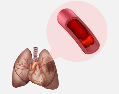
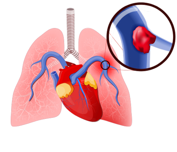
Pulmonary embolism is a blockage in one of the pulmonary arteries of the lungs. The most common reason for pulmonary embolism (PE) is a blood clot (thrombus) that travels to the lungs from other parts of the body (usually a leg) and becomes lodged blocking the blood vessel in the lung.

It is a medical emergency and immediate treatment can greatly increase the survival chances.

Pulmonary Embolism

Causes:

When a blood clot or in some cases, a clump of material causes a blockage in an artery of the lung, it leads to pulmonary embolism.

Clump of material other than blood clots include:

  • Fat (of marrow) from a long, broken bone
  • Collagen or other tissue
  • Part of a tumor
  • Air bubbles

In majority of the cases, multiple clots are involved but it is not necessary that all are involved in embolism. The affected part of the lung may die due to disruption of the blood supply. Moreover, it becomes difficult for the affected lung to supply oxygen to the rest of the body.

Signs and symptoms:

Its symptoms vary greatly depending on the size of the clots, the part of the lung affected, or pre-existing heart or lung disease.

Common signs and symptoms include:

  • Shortness of breath- it appears suddenly and becomes worse with exertion.
  • Chest pain- it is not relieved by rest and becomes worse upon cough or deep breath.
  • Cough- the cough may produce blood-tinged sputum.

Other signs and symptoms due to an underlying health condition or disease include:

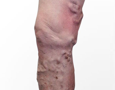
  • Leg pain or swelling
  • Discolored skin (cyanosis)
  • Fever
  • Profuse sweating
  • Rapid or abnormal heartbeat
  • Lightheadedness

Risk factors:

The risk factors are mostly similar to that of a patient with deep vein thrombosis (DVT) which include:

  • Longer hours of rest as seen in individuals who are hospitalized or even a long period of flight journey
  • Hypercoagulability (increased blood clotting potential) which may be caused by certain medications such as birth pills
  • Smoking
  • Cancer
  • Recent surgery
  • Pregnancy
  • Damage to the blood vessel wall

Diagnosis:

When a patient is suspected of pulmonary embolism or blood clots in the lung, then several crucial tests may be performed which include:

Pulse oximetry

It the simplest noninvasive way to measure the blood oxygen levels. A blood oxygen saturation level of 95 percent is abnormal.

Arterial blood gas

It is performed on a blood sample taken from an artery and the level of blood gases is evaluated. A partial pressure of oxygen less than 80mm Hg is abnormal.

Chest x-ray

It is done to rule out other diseases such as pneumonia or fluid in the lungs which may be responsible for the symptoms.

Ventilation-perfusion scan (VQ-scan)

It determines whether the person has experienced pulmonary embolism (PE) by evaluating both air flow and blood flow in the lungs. It also decides the probability of the PE which can be high, intermediate or low. A normal VQ scan suggests absence of PE.

Spiral computed tomography of the chest

It serves as an alternative to VQ scan. When PE is suspected, a contrast dye is injected through a vein to make the blood vessels stand out.

Pulmonary angiogram

When symptoms of PE tend to be non-conclusive even after a low or intermediate VQ scan or a normal spiral CT scan, then pulmonary angiogram is a definite test. It an invasive test that uses x-rays to reveal the blockage in the veins or arteries.

Echocardiogram

It is not particularly used to diagnose PE, but it evaluates the strain on the right side of the heart due to a large PE as well as some heart problems which may imitate PE. About 40 percent patients with PE have abnormalities of the right side of the heart, particularly the right ventricle.

Prevention:

It can accomplished by preventing the underlying conditions which cause PE, such as deep vein thrombosis and also by following the preventive measures which include:

  • Blood thinners or anticoagulants may be recommended for the patients with complications of cancer or after a stroke or heart attack.
  • Maintain a healthy weight.
  • Avoid smoking.
  • Consult a doctor about risk of clots before taking birth pills or hormone replacement therapy.
  • Exercise regularly for better blood circulation.
  • Drink adequate amount of fluids to stay hydrated.

Treatment:

Approximately 10% of the patients who develop pulmonary embolism die within the first hour without immediate treatment.

Immediate treatment greatly reduces the chances of death and shortens the stay in the hospital. The following treatment options are considered:

Compression stockings

These stocking are up to the knee and maintain a pressure on the legs to prevent blood clots. Sometimes they are also referred to as “support hose”.

Medications:

  • Blood thinners/anticoagulants- They are administered intravenously or in the form of pills depending on the patient’s condition and what works best for them. Blood thinners prevent further formation of blood clots but don’t dissolve the already existing blood clots. Most commonly used anticoagulants are heparin and warfarin. Warfarin is not recommended during pregnancy.
  • Thrombin inhibitors: It is recommended for people who can’t take heparin.
  • Thrombolytic medicines: It is recommended only during life threatening conditions after careful consideration as they can cause sudden bleeding.

Surgery:

Rarely a surgical intervention is used to remove a clot or to prevent it from reaching the lung. This procedure may include:.

  • Inferior vena cava filter– It is opted when the formation of blood clots can’t be resolved by the use of medication (blood thinners). In such a scenario, a filter is placed inside the inferior vena cava which prevents the blood clots to reach the lungs.
  • Catheter- In some emergency cases, a thin flexible tube is introduced into a vein in thigh or arm and it eventually reaches the affected blood vessel of the lung where the blood clot is dissolved by a medicine or it is removed.

Self-injectable at home:

Low molecular weight warfarin are available in self-injectable forms which include dalteparin, enoxaparin and tinzaparin, but it carries the risk of bleeding which has to be taken care of, while using such medications.

Book Online Consultaion


    Pulmonary Embolism Disease Blog

    DOWNLOAD PDF

    Subscribe the Hearty Life Blogs


      disease-condtions-deep_vein_thrombosis.png

      When a blood clot (thrombus) is formed in one or more of the deep veins of your body (usually in the legs), it is known as deep vein thrombosis (DVT). Sometimes this clot becomes detached from the inner walls of the blood vessel and travels to another part inside the body. DVT may develop due to an underlying medical condition particularly the one which affects the clotting of the blood.

      Causes and risk factors:

      It can be caused by the factors which prevent the normal circulation of the blood and it’s clotting, such as long periods of rest (during a particular medical condition), injury to the vein, surgery and certain medications.

      The risk factors are as follows:

      • Inherited blood clotting disorder
      • Prolonged bed rest
      • Surgery
      • Pregnancy
      • Contraceptives or hormone replacement therapy
      • Obesity
      • Smoking
      • Cancer
      • Heart failure
      • Inflammatory bowel disease
      • Age (usually older than 60 years but can also occur at any age)

      Complications:

      The following are the complications which may arise due to DVT:

      Pulmonary embolism: In Pulmonary embolism, a thrombus present in another part of your body usually a leg, travels to the blood vessel of the lung and blocks the blood circulation, leading to a life-threatening condition.

      Post-phlebetic syndrome (post thrombotic syndrome): set of signs and symptoms seen after a blood clot has formed.

      Symptoms:

      The person may be asymptomatic during the formation of the thrombus, however, during post thrombotic period when it has caused damage to the veins, the blood flow to the affected body part may be compromised. The following symptoms are observed:

      • Persistent swelling of the leg or arm (edema)
      • Leg pain or tenderness while standing
      • Skin discoloration in the affected arm or leg
      • Veins in the arm or leg appearing larger than normal

      Sometimes the development of a serious health complication such as pulmonary embolism is responsible for the symptoms in the DVT patients which are as follows:

      • Shortness of breath
      • Chest pain
      • Cough (blood may be coughed out as well)
      • Back pain
      • Profuse sweating
      • Lightheadedness
      • Blue nails or lips

      These symptoms appear suddenly, and an immediate medical attention is required.

      Diagnosis:

      The diagnosis is initiated by a thorough clinical examination and involves review of the patient’s medical history. Based upon these findings, the physician will categorize the patient into low or high risk DVT. Further tests may be considered to rule out other health problems and to confirm the diagnosis, which are as follows:

      • Blood tests: These are considered to evaluate any blood disorders that increase risk of DVT.
      • D- dimer test: In this test, the levels of a protein called D-dimer is assessed. In patients with DVT, high levels of this protein can be found in the blood.
      • Duplex ultrasonography: It is a non-invasive evaluation of the blood flow in the arteries and the veins of a patient. But this test is not preferred for a thrombus located very deep inside the body such as pelvis.
      • Venography: It is a specialized type of an x-ray procedure wherein a dye is injected into a vein of the foot, and the blood clot if present is detected.
      • Magnetic resonance imaging (MRI): It is performed in patients for whom ultrasound examination is inappropriate or unfeasible. It can even detect a thrombus present in the patient’s pelvis and the thigh.

      Prevention:

      A recent World Thrombosis Day Ipsos survey showed that there is less awareness of DVT in countries around the world. Thus, the first step in prevention is making the public aware of DVT. The preventive measures are different depending on the person’s condition who are at risk of developing DVT:

      During pregnancy

      In pregnant women, certain medication may be prescribed if she has risk factors such as likelihood of having a cesarean birth, bed rest, history of DVT, and inherited thrombophilia.

      During travel or in longer hours of sitting posture:

      • Drink lots of fluids.
      • Wear loose fitting clothes.
      • Walk and stretch at regular intervals.
      • Use special stockings to improve blood flow and to prevent blood clots but consult your health care provider before using it.

      Treatment:

      Treatment for DVT includes certain medicines and therapies, which are as follows:

      Medications:

      • Anticoagulants: They prevent further formation of blood clots but don’t dissolve the already existing ones. Most common anticoagulants used are heparin and warfarin. Warfarin is not recommended during pregnancy.
      • Thrombin inhibitors: These are recommended for people who can’t take heparin.
      • Thrombolytic medicines: These are recommended only during life threatening condition.

      Compression stockings:

      It may be recommended to prevent swelling and to avoid post-thrombotic symptoms in the patients.

      Surgical treatment:

      Surgical treatment may be performed when medications fail to dissolve the clots.

      Book Online Consultaion


        Deep Vein Thrombosis Blog

        DOWNLOAD PDF

        Subscribe the Hearty Life Blogs


          disease-condtions-thromboembolism.jpg

          Thromboembolism refers to a condition where a blood clot formed in the blood vessel unplugs itself and flows in the blood stream to block another blood vessel leading to obstruction of blood flow. Although clotting is a normal bodily function, if a clot occurs where it is not required, it may lead to serious problems.

          Types of Thromboembolism:

          The type of thromboembolism differs depending on the area in which the clot may appear. It can obstruct the blood flow in the veins, arteries, brain, gastrointestinal tract or kidneys.

          Thromboembolism can be further classified as below:

          Venous Thromboembolism:

          A venous thromboembolism is a thrombus (blood clot) formed in a vein. The blood flow in the veins is slow when compared to arteries which can increase the likelihood of the blood clots forming in these vessels.

          It can be categorized into three different types

          • Superficial Vein Thrombosis
            The clot may occur in a superficial vein, close to the surface of the body. Usually, it is not serious until it enters the deep veins.
          • Deep Vein Thrombosis (DVT)
            Deep vein thrombosis occurs within the deep veins, and mostly occurs within the legs.
          • Pulmonary embolism (PE)
            Pulmonary embolism is a medical emergency in which the thrombus blocks the blood vessel in the lungs.

          Renal Vein Thrombosis (RVT)

          A renal vein thrombosis is a thrombosis that occurs in the veins that drain blood away from the kidneys. These clots reduce the ability of the kidneys to clean and filter the blood.

          Arterial Thrombosis (atherothrombosis)

          Arterial thrombosis is much less common than venous thrombosis. It can have similar risks. Usually arterial thrombosis may lead to necrosis of the tissue.

          A thromboembolism in the coronary artery can cause a heart attack. If blood supply to the brain is disrupted, the patient may suffer a stroke.

          Symptoms:

          Signs and symptoms of venous thromboembolism include the following

          DVT

          • Pain in arm or leg, usually in the thigh or calf
          • Swollen leg or arm
          • Reddening or warming of the skin
          • Red streaks on the skin

          PE

          • Shortness of breath
          • Chest pain under your rib cage that can get worse when you take a deep breath
          • Rapid heart rate
          • Feeling lightheaded or passing out
          • Accentuated second heart sound

          Arterial Thrombosis

          • Muscle pain or spasm in the affected area
          • Numbness and tingling in the arm or leg
          • Pale color of the arm or leg (pallor)
          • Weakness of an arm or leg

          If the condition is severe, it may lead to blister formation and shedding of the skin, leading to tissue necrosis.

          Symptoms of a clot in an organ vary with the organ involved but may include:

          • Pain in the part of the body that is involved
          • Temporary decrease in organ function

          Causes:

          The blood clots can occur due to injury to a vein, consequence of a surgery, use of certain medications and lack of movement of the limbs.

          In the case of PE, the blood clot may block the blood vessels of the lungs. The affected portion of the lung may die due to loss of blood supply, the condition is called as pulmonary infraction, making it difficult to provide oxygen to the rest of the body.

          Occasionally, the blood vessels can be blocked by substances other than blood like collagen, part of a tumour, air bubbles etc.

          Risk factors:

          The common risk factors include

          • Inheriting a blood-clotting disorder
          • Prolonged immobility which may be due to bed rest or long trips
          • Injury or surgery
          • Pregnancy
          • Birth control pills or hormone replacement therapy
          • Being overweight or obese
          • Smoking
          • Cancer
          • Heart failure
          • Inflammatory bowel disease
          • Family history of deep vein thrombosis or pulmonary embolism

          Diagnosis:

          The diagnosis of thromboembolism includes the following tests which could be considered depending on the type:

          D-dimer: The blood sample is tested for the presence of D-dimer which is a marker for the presence of blood clots.

          Duplex Ultrasound: This is an imaging test in which the presence of clots is identified using ultrasound waves.

          Pulse oximetry: In this test, a sensor attached on the end of the finger of the patient helps to measure the level of oxygen in the blood.

          Arterial blood gas: The blood drawn from the artery is checked for oxygen levels present in it.

          Chest X-ray: This test may not be useful in finding the presence of clots, but can help to rule out a clot.

          Other diagnostic tests may include ELISA, pulmonary angiography, venography, echocardiography, helical computed tomography of pulmonary vessels etc.

          Treatment:

          Treatment of thromboembolism includes:

          Blood thinners: These are anticoagulant drugs which prevent formation of new clots while the body works to break up the previous clots present. They include heparin, low-molecular-weight heparin, apixaban, edoxaban, rivaroxaban, and warfarin.

          Clot-busting drugs: These are intravenous injections to dissolve the clots in the case of life-threatening situations. They include drugs belonging to the class of tissue plasminogen activators.

          Surgical and other procedures:

          • Removal of clot: Surgery is considered only if the condition is life threatening. The clot is removed using a catheter threaded through the blood vessel.
          • Vein filter: A catheter can also be used to position a filter in the inferior vena cava which helps to stop the clots from being carried into the lungs.

          Prevention:

          If the patient is at a risk of the condition, the following preventive measures may be considered to reduce the occurrence:

          • Use of blood thinners
          • Use of compression stockings
          • Use of intermittent pneumatic compression devices that automatically squeeze the legs to keep blood flowing

          Other preventive measures include

          • Regular exercise
          • Maintaining healthy weight
          • Cessation of smoking

          Consideration during travel include

          • Walk every 1 to 2 hours
          • Stretch legs and move around on the seat
          • Drink lots of fluids
          • Avoid smoking and consumption of alcohol before journey

          Book Online Consultaion


            Thromboembolism Blog

            DOWNLOAD PDF

            Subscribe the Hearty Life Blogs


              disease-condtions-varicose_veins.jpg

              Varicose veins are the twisted, swollen and bulging veins that appear in blue, red and skin color, just beneath the surface of the skin. They occur mostly in the legs and feet, causing aching pain and discomfort in the legs.

              Larger varicose veins appear rope-like and make the skin to bulge out. Inner varicose veins usually do not appear, but cause aching pain and swelling in the legs. Spider veins are the type of varicose veins which are most common and occur superficially, surrounded by the patches of flooded capillaries.

              Causes:

              Arteries have a thick layer of muscle or tissue to supply oxygenated blood to different parts of the body. Veins carry blood back to the heart and it should work against the gravity, so they rely on surrounding muscles pressure. Valves in the veins open to move the blood towards the heart and then close to prevent the back flow of blood.

              Any damage/abnormality in the functioning of the valves will lead to pooling up of blood in the vessels, causing them to stretch and twist, resulting in varicosity. It mostly occurs due to prolonged standing and walking, which builds up pressure in the veins of the lower legs.

              Signs and Symptoms:

              • Dark purple or blue color veins
              • Twisted and bulging veins that appear on the surface of the skin
              • Aching pain and heaviness in the legs
              • Burning, throbbing, muscle cramping and swelling in the lower legs
              • Worsened pain after sitting or standing for a long time
              • Itching around affected veins
              • Skin discoloration around a varicose vein
              • Web-like reddish blue varicose veins called spider veins

              Risk factors:

              The factors that increase the risk of developing varicose veins are:

              • Age: As the age increases, the functioning of the valves in the veins may be damaged, resulting in pooling up of blood in the lower leg veins, leading to varicose veins.
              • Gender: Hormonal changes in women during pregnancy, premenstruation and menopause will relax the vein walls, which increases the risk for varicose veins. Use of hormonal birth control pills also increases the risk.
              • Pregnancy: Blood volume increases during pregnancy to support the developing fetus, but can also produce a side effect of enlarged veins.
              • Family history: If any family member has varicose veins or deep vein thrombosis, it increases the risk.
              • Obesity: Being overweight will increase the pressure in the veins of lower legs.
              • Standing or sitting for long periods of time will reduce the free flow of blood in the blood vessels and results in clot formation.

              Complications:

              • Ulcers: Skin ulcers may form near the ankle, which is an indication of vascular disease.
              • Blood clots: Clots may occur in the veins deep inside the legs, causing swelling and leg pain. The condition is referred as thrombophlebitis.
              • Bleeding: Sometimes, the superficial veins may burst, causing bleeding.

              Diagnosis:

              A physical examination is performed, and the patient is enquired about the nature of leg pain diagnose varicose veins. The family medical history of varicose veins and information about their daily lifestyle activities is gathered. Certain tests may be needed to confirm the diagnosis of varicose veins, and include:

              • Ultrasound test: This is a non-invasive test, which uses sound waves to create the images of internal organs of the body. This test helps to identify the blood flow in the veins and also identify the clots and weakened or leaky valves in the veins.
              • Venogram: This test is a type of angiogram, which uses contrast dye to clearly view the blood flow through the veins, mainly if physician suspects the presence of large clots in the veins.

              Treatment:

              Initially, after the diagnosis of varicose veins, the doctor would recommend some lifestyle modifications that can relieve pain and reduce worsening of the condition, such as:

              • Perform exercises
              • Elevate the legs.
              • Avoid tight fitting clothes that can prevent the free circulation
              • Avoid long periods of standing or sitting

              Compression stockings: It will compress the leg veins and muscles and improve the blood flow efficiently, without forming any clots in the veins. The stockings are available in different sizes and lengths and can be bought without prescription in the local pharmacy.

              Sclerotherapy: This procedure is mainly used for severe varicose veins, and involves injecting a chemical solution near the varicose veins, which helps in closing and scarring of the veins.

              Laser treatment: This technique uses bursts of light reflected onto the vein, which makes the vein slowly fade and disappear. This test is mainly used to treat smaller varicose veins.

              Catheterization: This procedure uses a thin, flexible tube (catheter) that is inserted into the enlarged vein, which uses radiofrequency or laser energy, mainly to collapse the larger varicose veins.

              High ligation and vein stripping: Veins are tied off before it joins a deep vein and then it is removed through an incision.

              Ambulatory phlebectomy: Series of small punctures are made in the skin under general anaesthesia, to remove the smaller varicose veins.

              Endoscopic vein surgery: A thin tube attached with a camera is inserted into the vein to view and close the varicose veins and then removing them by making small incisions on the skin, under general anaesthesia.

              Prevention:

              Varicose veins cannot be prevented completely, but can be reduced by following some measures like:

              • Exercising regularly and losing extra weight
              • Eating a high-fiber and low-salt diet
              • Avoiding high heels and tight hosiery
              • Elevating the legs.
              • Changing the position regularly, when sitting or standing for long periods

              Book Online Consultaion


                Varicose Veins Blog

                DOWNLOAD PDF

                Subscribe the Hearty Life Blogs


                  +91 95424 75650

                  Call us now if you are in a medical emergency need, we will reply swiftly and provide you with a medical aid.


                  +91 95424 75650


                  Call us now if you are in a medical emergency need, we will reply swiftly and provide you with a medical aid.


                  Dr. Raghu | Heart Specialist in Hyderabad
                  Yashoda Hospitals, Sardar Patel Rd, behind Hari Hara Kala Bhavan, Kummari Guda, Shivaji Nagar, Secunderabad, Telangana 500003


                  Copyright © 2023, Dr. Raghu. All rights reserved.

                  [gtranslate]