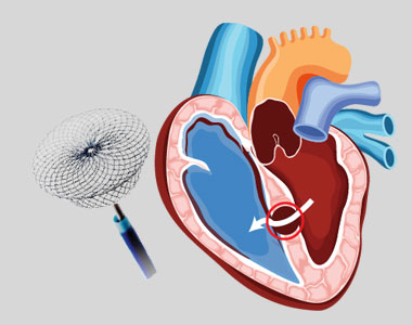
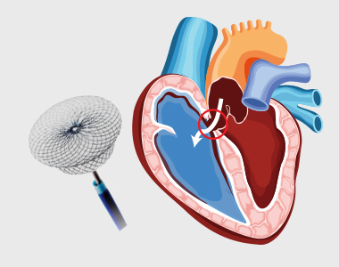
Patent Ductus Arteriosus (PDA) is a congenital heart condition in which the opening between the pulmonary artery and the aorta, persists after its normal closure time. The ductus arteriosus is a small connection between the two major blood vessels in the fetal heart, and it naturally closes shortly after birth. When it fails to close, it’s called patent ductus arteriosus. A PDA may allow the oxygenated blood to mix with the deoxygenated blood, compromising the heart and lung function. The larger PDA results in increased workload of heart, and also carries a risk of bacterial infection. PDA can be closed by inserting a device through the blood vessel in groin, a non-surgical method called percutaneous transcatheter approach. This method is considered only if the child is Considerations in adults include: PDA closure is a low risk procedure, but the common risks include:
What are the treatment options for PDA?
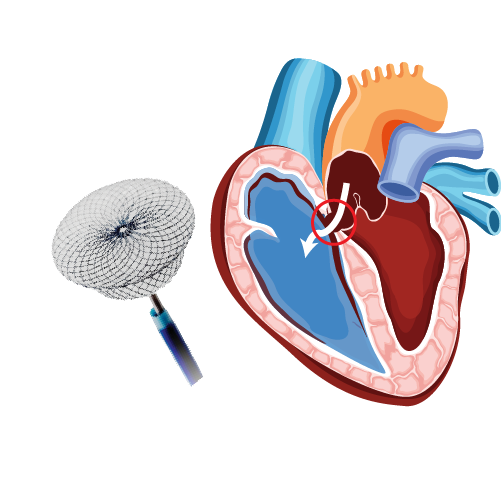
What is Device Closure for Patent Ductus Arteriosus (PDA)?
Who is eligible for this treatment?
What are the risks associated with the procedure?
What happens during the procedure?
What care should be taken post-procedure?
BOOK AN APPOINTMENT

Dr. RAGHU
Cardiology Coronary, Vascular and
Structural InterventionsConditions & Diseases
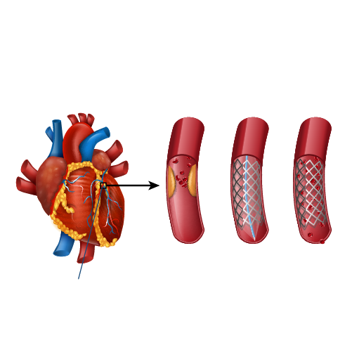
Angioplasty
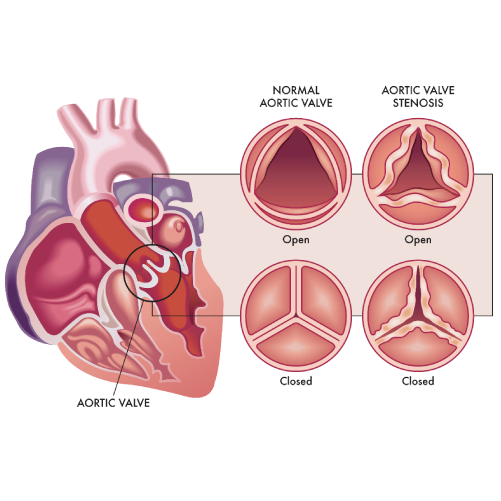
Aortic Stenosis
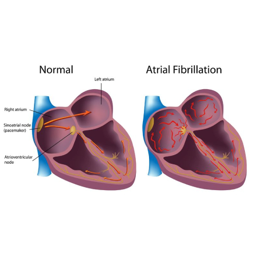
Atrial Fibrillation

Atrial Septal Defect
Uncategorized / Dr Raghu
Patent Ductus Arteriosus (PDA) is a congenital heart condition in which the opening between the pulmonary artery and the aorta, persists after its normal closure time. The ductus arteriosus is a small connection between the two major blood vessels in the fetal heart, and it naturally closes shortly after birth. When it fails to close, it’s called patent ductus arteriosus. A PDA may allow the oxygenated blood to mix with the deoxygenated blood, compromising the heart and lung function. The larger PDA results in increased workload of heart, and also carries a risk of bacterial infection. PDA can be closed by inserting a device through the blood vessel in groin, a non-surgical method called percutaneous transcatheter approach. This method is considered only if the child is Considerations in adults include: PDA closure is a low risk procedure, but the common risks include:

What are the treatment options for PDA?
What is Device Closure for Patent Ductus Arteriosus (PDA)?
Who is eligible for this treatment?
What are the risks associated with the procedure?
What happens during the procedure?
What care should be taken post-procedure?
BOOK AN APPOINTMENT
An atrial septum is a muscular wall that separates the upper chambers of the heart called atria. An ASD is a common congenital heart disease where the septum is not formed properly producing a left-to-right shunt, which leads to mixing of oxygenated and deoxygenated blood. This causes pulmonary hypertension and right heart enlargement. Small atrial defects do not need any treatment and close on its own. Even in adulthood small ASDs may remain asymptomatic. Some large defects that persists in adulthood may become symptomatic and need closure. The ASDs can be closed by: Percutaneous device closure is the preferred treatment for certain defects type. A noninvasive procedure known as percutaneous transcatheter approach is considered depending on the size and severity of the defect. Moderate to large-sized ASD along with pulmonary hypertension requires to be closed. The procedure is performed by inserting a special closure device either folded or attached to a catheter into the vein of the leg and is advanced to the heart through the defect, which closes the hole by a special mechanism. The success rate of the procedure is about 95%. But the risks involved, and their estimated incidence of occurrence include: Patients with small ASD may not develop any complications, but large-sized defects may lead to serious complications which demands surgery and prolonged hospitalization. A detailed diagnosis of the defect should be performed which includes transthoracic and transoesophageal echocardiogram used to assess the size, location and the suitability of the procedure.

What are the treatment options for ASD?
What is Percutaneous Closure of Atrial Septal Defect (ASD)?
Are there risks associated with the procedure?
What is the pre-procedure work-up?
How should I prepare for the procedure?
Are there any specific instructions about medications?
What happens during the procedure?
What care should be taken after the procedure?
BOOK AN APPOINTMENT
Dr. RAGHU
Cardiology Coronary, Vascular and
Structural InterventionsConditions & Diseases
Angioplasty
Aortic Stenosis
Atrial Fibrillation
Atrial Septal Defect
Copyright © 2023, Dr. Raghu. All rights reserved.
+91 95424 75650

+91 95424 75650