artery disease / Dr Raghu

disease-condtions-critical_limb_ischemia.png

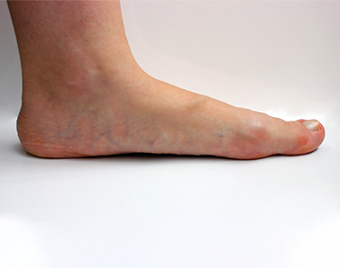
Critical limb ischemia occurs due to is a severe blockage of the arteries which leads to reduced blood flow to the extremities (hands, feet and legs). It results in severe pain and even skin ulcers or sores.

Causes:

Critical limb ischemia (CLI) is an advanced stage of peripheral artery disease (PAD) which results from progressive thickening of an artery’s lining caused by atherosclerosis (buildup of plaque). This eventually leads to narrowing of the artery which reduces normal blood circulation to the extremities.

Symptoms:

The early symptoms of ischemia of the limbs which can progress to critical limb ischemia involves pain, burning or cramping in the muscles of the limbs, usually after a physical activity or exercise which goes away with rest.

The following are the symptoms which may be indicative of critical limb ischemia:

  • Severe pain in the limbs or the legs when at rest
  • A noticeable low temperature of the lower leg or foot when compared to rest of the body
  • Toe or foot sores, infections or ulcers which heal slowly
  • Gangrene
  • Shiny, smooth, dry skin in the legs or feet
  • Thickening of the toenails
  • Absent or diminished pulse in the legs or the feet

Risk factors:

The risk factors include the following:

  • Age (over 60 years and post-menopausal women)
  • Smoking
  • Diabetes
  • Obesity
  • Sedentary lifestyle
  • High cholesterol
  • High blood pressure
  • Family history of vascular disease

Diagnosis:

Diagnosis is dependent on the location of ischemia. A thorough clinical examination is done as the symptoms are the first hint of this severe condition.

The following are the diagnostic methods which may be considered:

  • Ankle brachial index test: It is suggested when the ischemia is in the lower extremities of the body. It is a noninvasive procedure and helps evaluate the blood pressure in the legs.
  • Duplex ultrasound scanning: It is the most effective non-invasive scanning method of the arteries.
  • Magnetic resonance arteriography (MRA): The blood vessels are visualized based on magnetic resonance which enables the evaluation of stenosis.
  • Arteriogram: It is a contrast-enhanced x-ray of the arteries to help determine stenosis.

Prevention:

The following measures may help prevent peripheral artery disease:

  • Maintain a healthy active lifestyle.
  • Avoid or quit smoking.
  • Exercise regularly.
  • Maintain a healthy diet of low fat and low cholesterol.
  • Control blood sugar and blood pressure.
  • Manage stress.

Treatment:

Immediate treatment is required to reestablish the blood flow to the affected areas. The goal of the treatment should be to reduce pain and to improve blood flow to prevent amputation of the leg.

Treatment options include:

Medications
Prescribed medicines are aimed to prevent further progression of the disease and to reduce the effect of the factors that contribute to the risk factors involved. Medications that prevent clots or infections may also be prescribed.

Endovascular treatment

These are the least invasive methods which involves usage of a catheter. Angioplasty may be recommended to open the blockages and improve the blood circulation to the affected part of the limb. Laser atherectomy is a method in which laser is used to vaporize small bits of the plaque, followed by a catheter with rotating blade that physically removes the plaque from the artery.

Arterial surgery

It is recommended when the arterial endovascular treatment is not favorable. In this procedure, the diseased arterial part is removed or bypassed with a vein from the patient or with a synthetic graft.

Amputation

Amputation of the affected part is done as the last resort, and may be needed in about 25 percent of the CLI cases.

Book Online Consultaion


    Diabetic Foot and Critical Limb Ischemia Blog

    DOWNLOAD PDF

    Subscribe the Hearty Life Blogs


      disease-condtions-coronary_artery_disease.jpg


      Coronary artery disease (CAD) is the most common type of heart disease, in which the coronary arteries ( blood vessels that supplies oxygen and nutrients to the heart muscle) become rigid and narrowed due to buildup of plaque (deposition of cholesterol) in the inner walls of the heart. This restricts the flow of oxygen rich blood to the heart muscle, which may result in angina (chest pain) and shortness of breath. If the plaque ruptures or due to a complete blockage in the artery, it may result in heart attack or sudden cardiac death.

      Causes:

      Build of cholesterol plaques within the blood vessels is termed atherosclerosis.

      The plaque makes the inner walls of the artery sticky, so that the inflammatory cells, lipoproteins, and calcium travelling through the blood stream and adhere to the plaque. The plaque gradually enlarges, narrowing the blood vessel lumen, restricting blood flow to the heart muscle and thus reducing the supply of oxygen and nutrients to the heart.

      Signs and symptoms:

      • Angina (chest pain) – on exertion and in severe cases even at rest
      • Shortness of breath – on exertion and in severe cases even at rest
      • Palpitations (irregular heartbeats or skipped beats)
      • Tachycardia (fast heartbeat)
      • Weakness or dizziness
      • Nausea
      • Sweating
      • Fatigue, mainly after exercise or any activity

      Risk factors:

      • Age: Risk for damaged or narrowed arteries increases with age.
      • Gender: Men are at greater risk of developing CAD. In women, risk increases after menopause.
      • Family history: Risk increases if any family member has coronary artery disease.
      • Smoking: People who have the habit of smoking and who are exposed to smoke are at increased risk of CAD.
      • High blood pressure: It results in the narrowing and hardening of the walls of arteries.
      • High blood cholesterol levels: It can increase the risk of atherosclerosis.
      • Diabetes: High degree of association – 2 to 4-fold greater risk
      • Obesity: Overweight increases the risk of CAD.
      • Life style activities: Reduced physical activity, high stress and unhealthy diet also contribute to the development of CAD
      • Mental Stress: Responsible for young heart attacks Other possible risk factors, according to research, include:
      • Sleep apnea: It is a condition in which the person may experience intermittent cessation of breathing during sleep, resulting in reduced oxygen levels in the blood. Sleep apnea may cause an increase in the blood pressure and high strain on the heart, leading to CAD.
      • Increased C-reactive protein (CRP) levels in blood: CRP levels are increased mostly if there is any inflammation in the body. It is a risk factor for CAD.
      • High triglycerides (a type of lipid).
      • High homocysteine (amino acid) levels.
      • Preeclampsia (high blood pressure during pregnancy).
      • Alcohol use can lead to heart muscle damage.
      • Autoimmune diseases such as Rheumatoid arthritis and Lupus.

      Complications:

      If left untreated, CAD can lead to life threatening complications such as:

      • Angina:  Due to narrowed blood vessels, the heart muscles receive inadequate oxygen laden blood, causing chest pain or shortness of breath.
      • Heart attack: The plaque may rupture forming a clot and blocking the blood flow to the heart muscle.
      • Heart failure: Due to reduced oxygen and nutrient rich blood flow, the heart is unable to pump enough blood, thus increasing its workload. Over time, it may lead to weakening of heart muscle, also called heart failure.
      • Arrhythmia: Reduced amount of blood supply may lead to abnormal heart rhythms
      • Sudden cardiac death

      Diagnosis:

      The diagnostic work up includes a physical examination, in addition to medical history, family history, risk factors and any signs and symptoms of CAD. Further diagnostic tests may be recommended:

      • Electrocardiogram (ECG): It records the heart’s electrical activity and helps to determine the blood flow in the heart as well as heart muscle stress.
      • Echocardiogram: It uses sound waves to produce clear images of the heart and helps to identify the damaged heart muscles and valves.
      • Stress test: The test involves performing any high intensity activity like walking fast on a treadmill or bicycling under medical supervision, and close monitoring of heart function during the physical activity. Symptoms, ECG, blood pressure and heart rhythm are closely monitored during the test. Based on the findings the probability of having significant coronary artery disease would be determined. This test has many limitations and fallacies.
      • Cardiac catheterization and angiogram: A special medication called contrast is injected into the coronary blood vessels using a thin, flexible tube called catheter. The catheter is inserted through a small incision at the groin or through the wrist to visualise the blood vessels supplying the heart.
      • Heart scan: Computerized tomography (CT) technologies are used to identify the calcium deposits in the blood vessel of the heart, which indicates CAD.

      Coronary Artery Disease Treatment:

      Lifestyle changes:

      • Quit smoking
      • Avoid processed foods and eat high protein and fiber rich diet
      • Exercise regularly and lose excess weight
      • Manage stress
      • Control diabetes and blood pressure

      Medications:

      The doctor may prescribe medications, which help in reducing symptoms and severity of disease such as:

      • Dyslipidemic agents such as statins, ezeimibe to lower the blood cholesterol levels
      • Aspirin, a blood thinner, to prevent blood clots’ formation that might obstruct the coronary arteries
      • Beta blockers decrease the heart rate and blood pressure and reduce the oxygen demands of the heart.
      • Calcium channel blockers to lower blood pressure and improve blood supply to the heart.
      • Trimetazidine and Ranolazine are anti-anginal drugs to reduce chest pain
      • Nitroglycerin to dilate the coronary arteries and reduce chest pain
      • Angiotensin-converting enzyme (ACE) inhibitors and angiotensin II receptor blockers (ARBs) to decrease blood pressure and improve heart efficiency.

      Surgical procedures:

      • Angioplasty and stent placement: It is also called as percutaneous coronary revascularization, and involves inserting a thin, flexible tube (catheter) into the narrowed coronary artery, followed by a wire with deflated balloon to the narrowed area. The balloon is inflated to widen the blood vessel; a stent may also be placed to keep the artery patent.
      • Coronary artery bypass surgery: A graft is created using any blood vessel in the body, to bypass the blocked coronary arteries.

      Prevention:

      To maintain healthy arteries and to prevent coronary artery disease, some measures need to be taken such as lifestyle modifications which includes avoiding smoking and alcohol use, exercising regularly to maintain healthy weight, controlling sugar and cholesterol levels in blood and also eating a healthy diet.

      Book Online Consultaion


        Coronary Artery Disease Blog

        DOWNLOAD PDF

        Subscribe the Hearty Life Blogs


          +91 95424 75650

          Call us now if you are in a medical emergency need, we will reply swiftly and provide you with a medical aid.


          +91 95424 75650


          Call us now if you are in a medical emergency need, we will reply swiftly and provide you with a medical aid.


          Dr. Raghu | Heart Specialist in Hyderabad
          Yashoda Hospitals, Sardar Patel Rd, behind Hari Hara Kala Bhavan, Kummari Guda, Shivaji Nagar, Secunderabad, Telangana 500003


          Copyright © 2023, Dr. Raghu. All rights reserved.

          [gtranslate]