coronary arteries / Dr Raghu

New-Project-3.jpg

What Is Coronary Angiography?

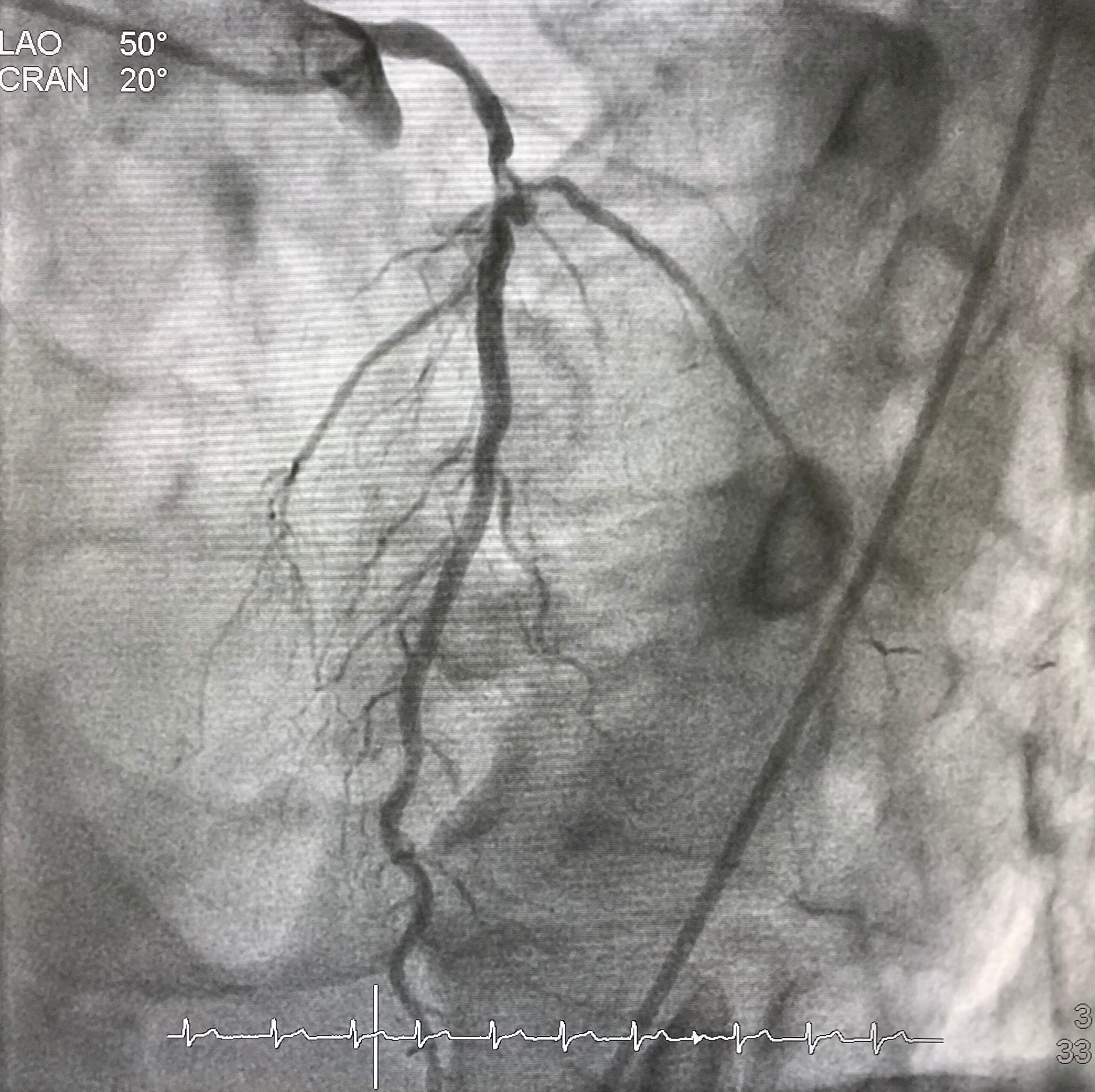
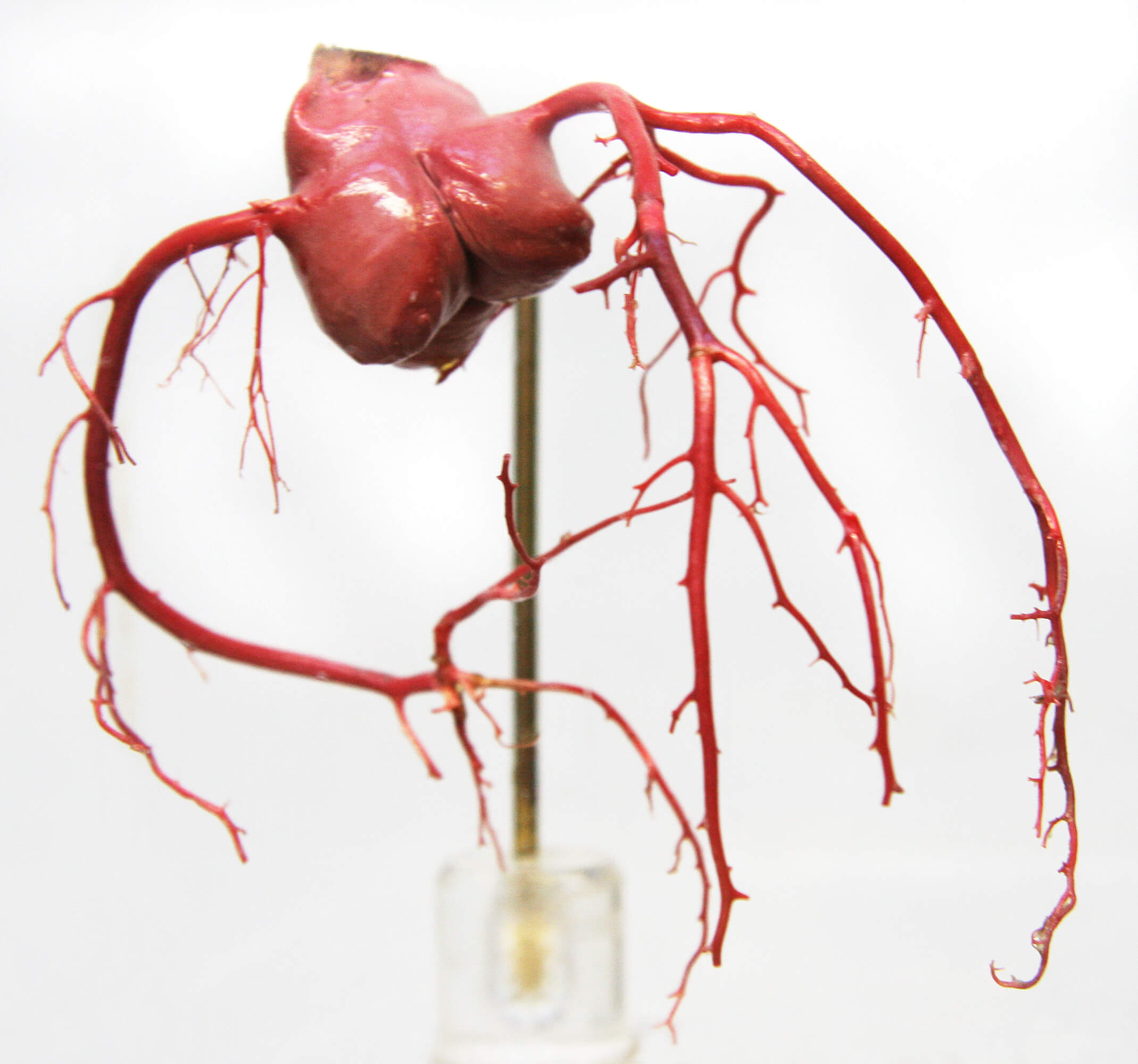
Coronary angiography is a procedure that uses X-rays to visualize and inspect arteries. It shows if there are any blocked arteries and how well your heart muscle is working.

During this procedure, a catheter is inserted into an artery in your arm or groin and advanced into one of your coronary arteries. Thereafter, contrast dye is injected into the coronary artery to make it visible on X-ray images.

The procedure can help identify blockages in the heart’s blood vessels and guide treatment decisions for patients at risk of developing heart disease or having a heart attack due to narrowed or blocked blood vessels that supply oxygenated blood to various organs.

When Is Coronary Angiography Performed?

Coronary angiography is typically performed if you have chest pain or other symptoms that suggest the presence of heart disease. If you have had a heart attack or have been diagnosed with coronary artery disease. It can be used in conjunction with an exercise stress test.

How Is Coronary Angiography Done?

If you’re scheduled for coronary angiography, here’s what you can expect:

  • The doctor will give you a sedative, usually in the form of an injection, at the start of your procedure.
  • They’ll insert a catheter into one of your arteries, either in your groin or wrist, and guide it through your blood vessels to reach your heart.
  • They’ll inject a special dye (contrast agent) into the coronary arteries that supply blood to your heart muscle so that they can see them clearly on X-rays taken after injecting this contrast agent.

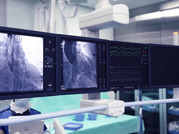
Where Is Coronary Angiography Performed?

Coronary angiography is performed in a cath lab (catheterization laboratory). The cath lab is a room with special equipment for performing coronary angiography and other procedures that entail inserting a long, thin tube (called a catheter) into the blood vessels of your heart.

Why Is Coronary Angiography So Common Nowadays?

Advancements in medical science have made coronary angiography more accessible to patients. The procedure has become simple and the risk has reduced significantly. Also, unhealthy diets and lifestyle choices put more people at risk of developing cardiac ailments. That’s why coronary angiography is commonly performed now-a-days.

What are the risks involved in Coronary angiography?

In expert hands coronary angiography is a near-zero risk procedure. The risk of complications can be broadly categorized into:

Less severe complications

  • bleeding under the skin at the wound site (haematoma) – this should improve after a few days, but contact your Cardiologist if you’re concerned. Application of ice packs would be helpful.
  • bruising – it’s common to have a bruise in your groin or arm for a few weeks. Application of ice packs would be helpful.
  • allergy to the contrast dye used, causing symptoms such as a rash and a headache – this is uncommon, but you should discuss any allergies with your cardiologist before having the procedure

Severe complications

The chance for developing a serious complication during coronary angiogram is 1 in 1000. People with serious underlying heart problems are most at risk. Discuss with your cardiologist about the risks before the procedure.

  • damage to the artery in the arm or groin in which the catheter was inserted, with the blood supply to the limb possibly being affected 
  • heart attack – a serious medical emergency where the heart’s blood supply is suddenly blocked 
  • stroke – a serious medical condition that occurs when the blood supply to the brain is interrupted 
  • damage to the kidneys caused by the contrast dye 
  • tissue damage caused by X-ray radiation if the procedure is prolonged 
  • death

In Conclusion

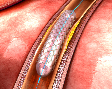
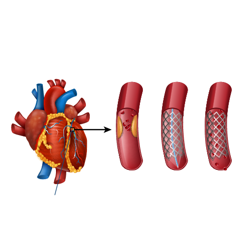
Coronary angiography is used to diagnose and treat heart diseases, before cardiac surgery, angioplasty-stent procedures as well as other conditions such as aneurysms in blood vessels. It helps doctors identify underlying causes of heart failure and determine the proper course of treatment.

Dr. C Raghu is a renowned cardiologist with decades of experience in interventional cardiology. He is one of the pioneers of trans-radial procedures in India. Consult him if someone is in need for coronary angiogram.

Book Online Consultaion


    Coronary Angiography Blog

    DOWNLOAD PDF

    Subscribe the Hearty Life Blogs


      Coronary-Bifurcation-Lesions.jpg

      Coronary arteries are the vessels which supply blood (oxygen and nutrients) to the heart; this supply may be disrupted due to buildup of cholesterol and fatty deposits, called plaques, on the inner walls of the arteries (atherosclerosis) causing narrowing of the vessel lumen.

      Coronary lesions branch out as they progress to supply various cardiac structures. A bifurcation lesion is a stenosis or abnormal narrowing of greater than 50% that occurs in a coronary artery at the origin of the side branch or adjacent to the origin of the side branch. Branching points a.k.a bifurcation points in the coronary arteries are prone to develop atherosclerosis as the shear stress is higher at the branch points.

      The figure shows how a bifurcation stenosis appears schematically as well as on angiography.

      bifurcation lesion


      Why bifurcation lesions are important?

      A vast majority of coronary arteries obstructions happen at the branching point. About 15-20% of patients of referrals for bypass surgery are because of the presence of coronary narrowing at bifurcation location. They are considered a tough lesion to treat by angioplasty and stent.

      How are bifurcation lesions diagnosed?

      Bifurcation lesions are identified on coronary angiography. Only lesions affecting side branch blood vessels more than 2.25 mm is considered important and called bifurcation lesions. The main vessel that gives branches is called main vessel and the branches that originate from it are called side branches. The point at the division of the main vessel and side branch is called a bifurcation point.

      How is a bifurcation lesion unique?

      Bifurcation lesions can involve the coronary artery either before or after the bifurcation point. In addition, side branches could be either diseased or free from disease. Based on the location and severity of the cholesterol plaque at the bifurcation point, sub types of bifurcation lesions have been identified.

      The challenge of performing angioplasty and stent for a bifurcation stenosis lies in the ability to preserve the main vessel and the side branch. Stents being metallic tubes risk occluding the side branch when placed across the side branch. This apprehension of losing the side branch, is one of the primary reasons for patients to be sent for bypass surgery instead of angioplasty and stent.

      Coronary-bifurication

      Can angioplasty and stent be performed for a bifurcation lesion?

      Angioplasty and stent can be eminently performed for a bifurcation stenosis. Over the last 2 decades numerous techniques in stent implantation have been developed to treat the bifurcation lesions. Some of these techniques include:

      • Provisional T stent technique
      • Tap technique
      • Double kiss crush technique
      • Culotte technique

      Advent of these techniques enabled experienced operators to perform angioplasty and stent successfully with chances of repeat narrowing seen in 3-6% patients at the site of stenting. Risk of losing a side branch after stenting is reduced to 1% once the new techniques are employed.

      Dr. C Raghu is an expert in bifurcation lesion angioplasty and the center where he has been trained, ICPS Paris France is considered the “Mecca” for bifurcation lesion angioplasty techniques development. 

      How does a bifurcation lesion angioplasty differ from a normal angioplasty?

      Bifurcation lesion angioplasty, involves utilization of novel stent techniques mentioned above. In addition, angioplasty operator experience is a major factor in determining  outcomes. Choosing the “right” stent that allows side branch access, use of specialized technologies such as fractional flow reserve (FFR), expertise in deploying 2 stents especially the process of “stent crush” and the handling of two balloons through a single catheter – kissing balloon are key points for the best results in a stent procedure.

      What are the chances of side branch occlusion in a bifurcation lesion angioplasty?

      Contemporary bifurcation angioplasty using Intravascular imaging employing contemporary stenting techniques in the hands of expert bifurcation angioplasty operators carries a 99% success rate with a chance of losing side branch in 1% and repeat narrowing at the end of 9 months being 3-6%. These results are comparable and if not superior to bypass surgery as angioplasty entails a short hospital stay and fast recovery.

      English Video

      Book Online Consultaion


        Coronary Bifurcation Lesions Blog

        DOWNLOAD PDF

        Subscribe the Hearty Life Blogs


          cci-coronary_angioplasty.jpg


          Coronary arteries are the blood vessels that delivers oxygenated blood to the heart muscle. But, sometimes these blood vessels may get narrowed or blocked due to accumulation of cholesterol plaque. To open the blockage and restore the function of arteries, coronary angioplasty is performed.

          A coronary angioplasty involves the temporary insertion of a tiny balloon inside the artery to open the blockage. It is usually combined with the placement of a stent (a metal mesh tube) to widen the artery and prevent the further chance of narrowing.

          coronary angioplasty

          Why is coronary angioplasty done ?

          Coronary angioplasty is done to treat narrowing or blockage of the heart’s blood vessels:

          • during or after a heart attack.
          • Blood vessel narrowing leading to poor heart function.
          • For relief of chest pain (angina) due to reduced blood supply to heart.

          Risks of coronary angioplasty

          Usually, it is a safe procedure, but like all other procedures, it also carries certain risks like:

          Common Issues/Risks after angiography
          Bruising Common to have bruise at groin or arm Lasts for few weeks Not to be worried about
          Allergic reactions Usually related to contrast medicine used History of previous allergy – higher risk Present as rash and headache
          Bleeding at the site of entry (Hematoma) Usually subsides in a few days But in case swelling is increasing and painful contact your physician immediately


          Serious complications after coronary angiography Less than 1 in 1000 chance People with severe heart disease are at high risk Discuss with Cardiologist before procedure
          Heart attack A serious medical emergency where the heart’s blood supply is suddenly blocked
          Stroke A serious medical condition that occurs when the blood supply to the brain is interrupted
          Loss of blood supply to limb Damage to the artery in the arm or groin in which the catheter was inserted, with the blood supply to the limb possibly being affected
          Kidney injury Damage to the kidneys caused by the contrast dye
          Radiation injury Tissue damage caused by X-ray radiation if the procedure is prolonged

          What happens before the procedure?

          A physical examination and blood test are done to evaluate the overall health condition. The person needs to follow instructions provided before undergoing the procedure:

          1. Inform the doctor about:

          • Any allergies
          • The use of current medicines, including vitamin and mineral supplements
          • Any blood disorder
          • Any surgery you may had
          • The past and present medical condition

          2. Medicines:

          Tell the doctor if you are using blood thinners or any anti-diabetic medicines. The doctor will either advise you to stop or change the dose of the medication.

          3. Food and fluid restrictions:

          • Avoid heavy meals, such as meat, fried or fatty foods eight hours before the procedure. Take light foods such as toast and cereal.
          • Fast for six hours before the procedure.
          • Stay hydrated, drink plenty of fluid up to two hours before the procedure.

          In most of the cases, you will be discharged on the same day of the hospital, so ask someone to accompany you to the hospital.

          What happens during the procedure?

          Local anaesthesia would be administered to numb the area where the catheter would be inserted. Mostly, a catheter is inserted through the wrist, and sometimes through the groin. Under fluoroscopy (a type of X-ray), the catheter will be guided to the diseased artery. Then a special medicine (called contrast or dye) will be injected into the blood vessels supplying the heart. This will delineate the presence of obstruction to blood flow. 
          Then a wire is passed across the obstruction and a balloon is inflated at the site of obstruction to compress the obstructing plaque in the artery. The balloon expansion will compress the plaques into the wall of the blood vessel and improve the blood flow. Once the blood flow is improved, the catheter will be removed, and a stent would be placed to prevent the risk of further blockage. Finally, the catheter will be removed and the puncture site is sealed.

          What to expect after the procedure?

          The nurse will check the vitals of the patient. Depending on the condition, the doctor would advise you the hospital stay. You will be advised not to bend or cross the leg for a few days if the procedure is performed through the groin region. Additionally, some other tests like X-rays and an electrocardiogram (ECG) would be done to check the condition. Before discharge, the doctor will give you certain instructions regarding wound care, medications and lifestyle.

          Recovery after coronary angioplasty

          Recover quickly and keep your heart healthy by following these tips:

          • Quit smoking.
          • Limit the intake of alcohol.
          • Take the prescribed medicines.
          • Check the cholesterol levels regularly.
          • Maintain a healthy body weight.
          • Have a healthy and well-balanced diet.
          • Exercise regularly.

          Know your numbers

          It’s important to periodically monitor and maintain the following numbers to prevent future cardiac events

          • LDL cholesterol – less than 55 mg/dL
          • Blood pressure – less than 120/80 mm Hg
          • Blood sugar – HbA1C less than 7.0%

          Book Online Consultaion


            Download: Coronary Angioplasty Blog

            GET DOWNLOAD PDF NOW

            Subscribe the Hearty Life Blogs


              disease-condtions-coronary_artery_disease.jpg


              Coronary artery disease (CAD) is the most common type of heart disease, in which the coronary arteries ( blood vessels that supplies oxygen and nutrients to the heart muscle) become rigid and narrowed due to buildup of plaque (deposition of cholesterol) in the inner walls of the heart. This restricts the flow of oxygen rich blood to the heart muscle, which may result in angina (chest pain) and shortness of breath. If the plaque ruptures or due to a complete blockage in the artery, it may result in heart attack or sudden cardiac death.

              Causes:

              Build of cholesterol plaques within the blood vessels is termed atherosclerosis.

              The plaque makes the inner walls of the artery sticky, so that the inflammatory cells, lipoproteins, and calcium travelling through the blood stream and adhere to the plaque. The plaque gradually enlarges, narrowing the blood vessel lumen, restricting blood flow to the heart muscle and thus reducing the supply of oxygen and nutrients to the heart.

              Signs and symptoms:

              • Angina (chest pain) – on exertion and in severe cases even at rest
              • Shortness of breath – on exertion and in severe cases even at rest
              • Palpitations (irregular heartbeats or skipped beats)
              • Tachycardia (fast heartbeat)
              • Weakness or dizziness
              • Nausea
              • Sweating
              • Fatigue, mainly after exercise or any activity

              Risk factors:

              • Age: Risk for damaged or narrowed arteries increases with age.
              • Gender: Men are at greater risk of developing CAD. In women, risk increases after menopause.
              • Family history: Risk increases if any family member has coronary artery disease.
              • Smoking: People who have the habit of smoking and who are exposed to smoke are at increased risk of CAD.
              • High blood pressure: It results in the narrowing and hardening of the walls of arteries.
              • High blood cholesterol levels: It can increase the risk of atherosclerosis.
              • Diabetes: High degree of association – 2 to 4-fold greater risk
              • Obesity: Overweight increases the risk of CAD.
              • Life style activities: Reduced physical activity, high stress and unhealthy diet also contribute to the development of CAD
              • Mental Stress: Responsible for young heart attacks Other possible risk factors, according to research, include:
              • Sleep apnea: It is a condition in which the person may experience intermittent cessation of breathing during sleep, resulting in reduced oxygen levels in the blood. Sleep apnea may cause an increase in the blood pressure and high strain on the heart, leading to CAD.
              • Increased C-reactive protein (CRP) levels in blood: CRP levels are increased mostly if there is any inflammation in the body. It is a risk factor for CAD.
              • High triglycerides (a type of lipid).
              • High homocysteine (amino acid) levels.
              • Preeclampsia (high blood pressure during pregnancy).
              • Alcohol use can lead to heart muscle damage.
              • Autoimmune diseases such as Rheumatoid arthritis and Lupus.

              Complications:

              If left untreated, CAD can lead to life threatening complications such as:

              • Angina:  Due to narrowed blood vessels, the heart muscles receive inadequate oxygen laden blood, causing chest pain or shortness of breath.
              • Heart attack: The plaque may rupture forming a clot and blocking the blood flow to the heart muscle.
              • Heart failure: Due to reduced oxygen and nutrient rich blood flow, the heart is unable to pump enough blood, thus increasing its workload. Over time, it may lead to weakening of heart muscle, also called heart failure.
              • Arrhythmia: Reduced amount of blood supply may lead to abnormal heart rhythms
              • Sudden cardiac death

              Diagnosis:

              The diagnostic work up includes a physical examination, in addition to medical history, family history, risk factors and any signs and symptoms of CAD. Further diagnostic tests may be recommended:

              • Electrocardiogram (ECG): It records the heart’s electrical activity and helps to determine the blood flow in the heart as well as heart muscle stress.
              • Echocardiogram: It uses sound waves to produce clear images of the heart and helps to identify the damaged heart muscles and valves.
              • Stress test: The test involves performing any high intensity activity like walking fast on a treadmill or bicycling under medical supervision, and close monitoring of heart function during the physical activity. Symptoms, ECG, blood pressure and heart rhythm are closely monitored during the test. Based on the findings the probability of having significant coronary artery disease would be determined. This test has many limitations and fallacies.
              • Cardiac catheterization and angiogram: A special medication called contrast is injected into the coronary blood vessels using a thin, flexible tube called catheter. The catheter is inserted through a small incision at the groin or through the wrist to visualise the blood vessels supplying the heart.
              • Heart scan: Computerized tomography (CT) technologies are used to identify the calcium deposits in the blood vessel of the heart, which indicates CAD.

              Coronary Artery Disease Treatment:

              Lifestyle changes:

              • Quit smoking
              • Avoid processed foods and eat high protein and fiber rich diet
              • Exercise regularly and lose excess weight
              • Manage stress
              • Control diabetes and blood pressure

              Medications:

              The doctor may prescribe medications, which help in reducing symptoms and severity of disease such as:

              • Dyslipidemic agents such as statins, ezeimibe to lower the blood cholesterol levels
              • Aspirin, a blood thinner, to prevent blood clots’ formation that might obstruct the coronary arteries
              • Beta blockers decrease the heart rate and blood pressure and reduce the oxygen demands of the heart.
              • Calcium channel blockers to lower blood pressure and improve blood supply to the heart.
              • Trimetazidine and Ranolazine are anti-anginal drugs to reduce chest pain
              • Nitroglycerin to dilate the coronary arteries and reduce chest pain
              • Angiotensin-converting enzyme (ACE) inhibitors and angiotensin II receptor blockers (ARBs) to decrease blood pressure and improve heart efficiency.

              Surgical procedures:

              • Angioplasty and stent placement: It is also called as percutaneous coronary revascularization, and involves inserting a thin, flexible tube (catheter) into the narrowed coronary artery, followed by a wire with deflated balloon to the narrowed area. The balloon is inflated to widen the blood vessel; a stent may also be placed to keep the artery patent.
              • Coronary artery bypass surgery: A graft is created using any blood vessel in the body, to bypass the blocked coronary arteries.

              Prevention:

              To maintain healthy arteries and to prevent coronary artery disease, some measures need to be taken such as lifestyle modifications which includes avoiding smoking and alcohol use, exercising regularly to maintain healthy weight, controlling sugar and cholesterol levels in blood and also eating a healthy diet.

              Book Online Consultaion


                Coronary Artery Disease Blog

                DOWNLOAD PDF

                Subscribe the Hearty Life Blogs


                  +91 95424 75650

                  Call us now if you are in a medical emergency need, we will reply swiftly and provide you with a medical aid.


                  +91 95424 75650


                  Call us now if you are in a medical emergency need, we will reply swiftly and provide you with a medical aid.


                  Dr. Raghu | Heart Specialist in Hyderabad
                  Yashoda Hospitals, Sardar Patel Rd, behind Hari Hara Kala Bhavan, Kummari Guda, Shivaji Nagar, Secunderabad, Telangana 500003


                  Copyright © 2023, Dr. Raghu. All rights reserved.

                  [gtranslate]