Coronary artery disease / Dr Raghu

Symptom-of-congestive-heart-failure-2.jpg


There are many scenarios where you might want to see a doctor and find out whether you’ve developed heart failure. Perhaps you have a history of heart disease in the family and would like to assess your risk levels.

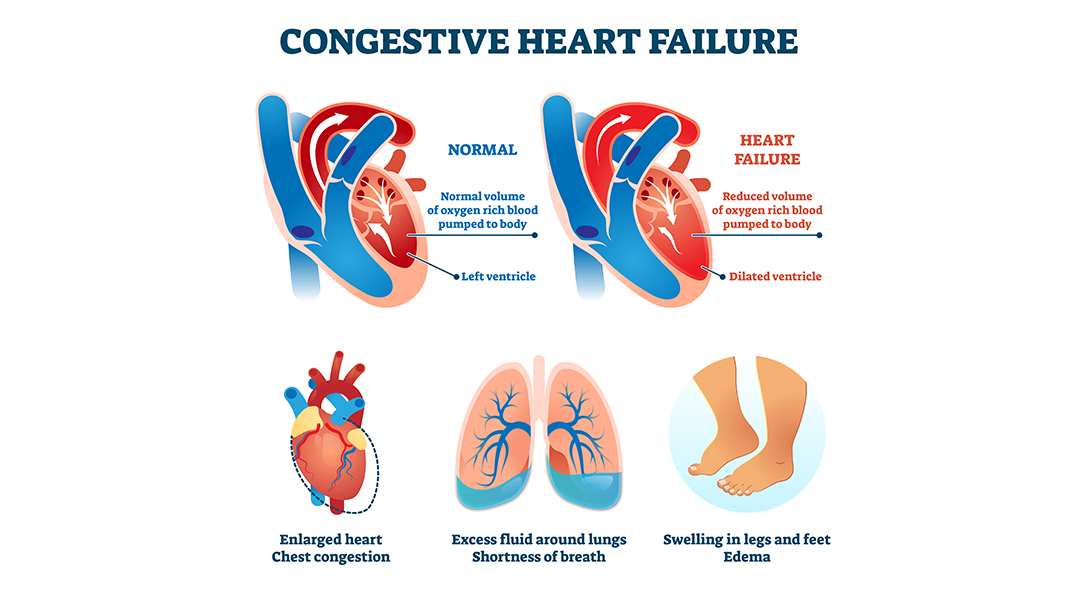
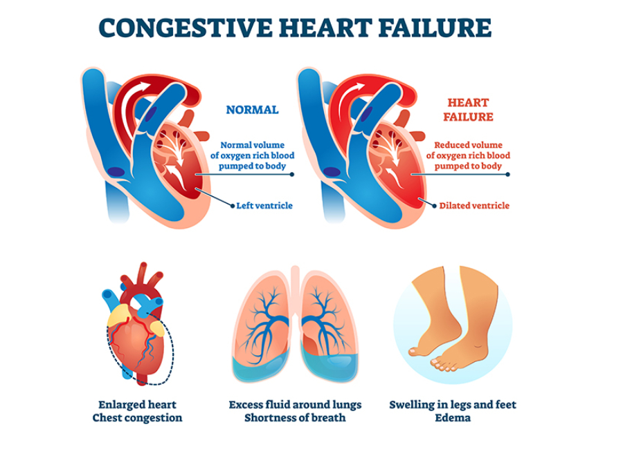
Or you might have developed symptoms, such as shortness of breath, chest pain, and swollen feet, that are indicative of heart failure. (Check out our previous article for a detailed glimpse of heart failure symptoms.)

Congestive Heart Failure

Or you might have completed an initial round of investigation and want a closer look at the root cause of heart failure. In any case, it’s essential to have a fair idea of the steps involved in diagnosing heart failure. Let’s take a look.

Family History and Medical Background

Diagnostic efforts for heart failure serve two primary purposes :

  • To determine the underlying cause
  • To assess the extent of the heart’s malfunction

The first thing a doctor will do is get a complete picture of your medical history. They’ll want to know the details of any symptoms you might have been experiencing. Also, they’ll ask about your diet and lifestyle, including your habits pertaining to exercise, smoking, and alcohol consumption.

Additionally, they’ll ask one or more of the following questions:

  • Do you have pre-existing conditions like high cholesterol levels, hypertension, diabetes, etc.?
  • Have you undergone treatments like chemotherapy?
  • Do you have a family history of cardiovascular diseases?

Your answers to these questions will give your doctor a better idea of your current physical condition.

Physical Examination

Next, the doctor will perform a thorough physical exam to analyze your heart activity. They’ll likely start by calculating your BMI and body fat percentage. Also, they’ll measure your vitals, including blood pressure and heart rate.

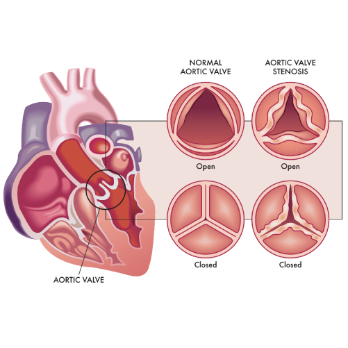
Additionally, they might use a stethoscope to identify abnormal heart sounds or murmurs that indicate a faulty heart valve. They’ll also watch out for soft noises or bruits to identify the narrowing of arteries.

They’ll examine your skin to see if it feels cold or looks discolored. They’ll also check your feet and abdomen for signs of fluid buildup. By the end of the physical exam, the doctor will have a better idea of your cardiovascular health.

Related : High Blood Pressure – Symptoms & Treatment

Diagnostic Tests

While a physical exam can indicate abnormal heart function, your doctor will likely run a few diagnostic tests to confirm the underlying cause of heart failure.

The most common tests include:

  • Blood tests, such as complete blood count, lipid panel, liver and kidney function tests, and a fasting glucose test
  • Brain natriuretic peptide (BNP) test to determine the risk of hospitalization and death due to heart failure
  • Chest X-ray to detect enlarged heart muscle or fluid buildup around the heart
  • 12-lead ECG to monitor the heart’s electrical activity and identify signs of a heart attack or irregular heartbeat
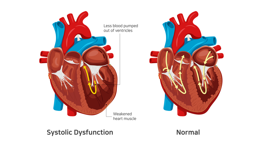
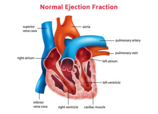
  • Echocardiography for a closer look at the heart’s chambers and pumping action in real time
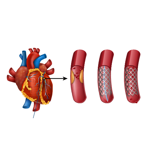
  • Coronary angiography to identify coronary artery disease

Other tests like radionuclide ventriculography, exercise testing, and endomyocardial biopsy may also be prescribed.

Related : What is Coronary Angiogram?

In Conclusion

Diagnosing heart failure involves a combination of physical examination, blood tests, and non-invasive procedures like X-rays and ECG. The key is to identify the underlying cause of heart failure, so that your doctor can decide the right course of treatment.

Dr. C Raghu is an experienced cardiologist who specializes in interventional cardiology. If you or anyone you know is at risk of developing heart failure, reach out to Dr. Raghu for a thorough diagnosis.

Book Online Consultaion


    Diagnosing Congestive Heart Failure Blog

    DOWNLOAD PDF

    Subscribe the Hearty Life Blogs


      Diastolic-heart-failure-img.jpg

      In our previous articles, we’ve discussed the symptoms of heart failure and the steps to diagnose the same. However, the course of treatment varies for every patient based on the type of heart failure they’ve developed.

      Depending on the part of the heart’s pumping cycle that’s been affected, there are two types of heart failure. In this blog, we’ll take a closer look at diastolic dysfunction and its symptoms.

      What Causes Diastolic Dysfunction?

      diastolic dysfunction

      The diastolic phase refers to the part of the heart’s pumping cycle when the ventricles (lower chambers) relax and let blood flow in from the atria (upper chambers). Diastolic dysfunction is a condition in which the ventricles don’t relax enough. That, in turn, prevents the normal amount of blood from entering the heart.

      Diastolic dysfunction is caused when the heart muscles become thicker and stiffer than usual. It’s more common in older women with hypertension and diabetes. If left untreated, it can lead to diastolic heart failure (also known as heart failure with preserved ejection fraction).

      Related : Understanding Congestive Heart Failure Symptoms

      What Does Preserved Ejection Fraction Mean?

      Ejection fraction refers to the volume of blood pumped out from the heart’s left ventricle with each contraction. For a healthy heart, the number falls in the range of 55% to 65%. A lower ejection fraction is one of the most common indicators of heart failure.

      However, it’s worth noting that many people with diastolic dysfunction have an ejection fraction of 50% or more (which is known as preserved ejection fraction). That means the left ventricle expels an adequate amount of oxygenated blood.

      However, the heart muscle doesn’t relax enough to let a sufficient quantity of blood in. That, in turn, causes the excess blood to back up in the lungs and results in fluid buildup in the feet and abdomen.

      How to differentiate systolic from diastolic dysfunction ?

      diastolic dysfunction

      In contrast to systolic dysfunction where the heart muscle is “weak”, in diastolic dysfunction the heart is “stiff”. This means that the heart is unable to pump blood out of the heart in systolic dysfunction whereas the heart is unable to accept further blood in diastolic dysfunction. Both conditions lead to congestion or fluid accumulation in various organs of the body. Differentiation of heart failure from systolic and diastolic dysfunction is not possible as both diseases present with similar symptoms. 

      Which conditions lead to Diastolic dysfunction?

      • Diastolic dysfunction appears consequent to uncontrolled or long-standing diabetes
      • Hypertension
      • Obesity as well as elderly people
      • Women and atrial fibrillation

      The best way to prevent and treat diastolic dysfunction is by effective control of the diseases mentioned above.

      Symptoms of Diastolic Dysfunction

      The most common symptom of diastolic dysfunction is congestion and shortness of breath due to the buildup of blood and fluid in the lungs. Breathing difficulties can get particularly worse during exertion or when lying.

      Other symptoms of diastolic dysfunction include:

      • Coughing and wheezing (due to lung congestion)
      • Loss of appetite and nausea (due to fluid buildup around the liver and in the stomach)
      • Swollen feet, legs, and abdomen (due to fluid accumulation)

      If you experience any of the given symptoms, it’s crucial to consult a doctor for a proper diagnosis.

      Treatment of Diastolic Dysfunction

      Treatment of diastolic dysfunction involves a combination of medications (diuretics or water pills) and lifestyle changes. In severe cases, a patient might need left ventricular assist devices or a heart transplant.

      Is Diastolic Dysfunction Serious?

      In the long run, diastolic dysfunction can lead to diastolic heart failure. That, in turn, increases your risk of hospitalization and death. Therefore, you should pay close attention to your symptoms and reach out to a doctor whenever you notice anything unusual.

      Dr. C Raghu is a renowned cardiologist who specializes in interventional cardiology. He has decades of experience in treating patients with different heart conditions. If you or anyone you know has developed symptoms like shortness of breath, swollen feet, loss of appetite, etc., contact Dr. Raghu to explore your treatment options.

      Book Online Consultaion


        What Are the Symptoms of Diastolic Dysfunction ? – Blog

        DOWNLOAD PDF

        Subscribe the Hearty Life Blogs


          Systolic-heart-failure-img.jpg

          Heart failure is a common condition with no known cure. However, proper treatment can control the disease progression and thus improve a patient’s quality of life and longevity. To decide the proper course of treatment, a doctor must first determine the type of heart failure a patient has developed.

          Depending on the part of the heart’s pumping cycle that’s been affected, heart failure can be of two types – diastolic and systolic. You can learn more about the symptoms, causes, treatment of diastolic dysfunction and differentiation from systolic dysfunction in our previous article.

          In this blog, we’ll delve deeper into systolic heart failure and understand its causes and symptoms.

          Systolic Heart Failure: A Closer Look

          Systolic Heart Failure

          Systolic heart failure occurs due to a problem in the heart’s contraction (or systolic) phase. It’s characterized by stretching and weakening of the left ventricular muscle, due to which the heart pumps out less oxygenated blood to the body.

          It’s also known as heart failure with reduced ejection infraction. As the condition worsens, it can also weaken the right ventricle and take a toll on its pumping power too.

          Related: What Are the Symptoms of Diastolic Dysfunction?

          Causes of Systolic Heart Failure

          Systolic heart failure is caused by underlying medical conditions that damage the left ventricle. The most common causes include :

          • Hypertension (the left ventricle has to use increased pressure to pump blood through the body)
          • Coronary artery disease (buildup of cholesterol in the arteries) – with or without a heart attack.
          • Dilated cardiomyopathy (weakening of the left ventricle due to an infection or long-term exposure to alcohol and narcotics)
          • Abnormal heart rhythm (also known as atrial fibrillation)
          • Previous heart attack

          Additionally, people who are older or have diabetes are at a higher risk of developing systolic heart failure.

          Related: Understanding Congestive Heart Failure Symptoms

          Symptoms of Systolic Heart Failure

          In systolic heart failure, an adequate amount of oxygen-rich blood doesn’t reach all organs. The most common indicator of the condition is a lower ejection fraction.

          It can result in the following symptoms:

          • Breathlessness – initially on exertion and in later stages even at rest or lying down. 
          • Swelling of feet, face, abdomen – due to fluid accumulation in various organs 
          • Engorged and pulsatile neck veins
          • Confusion (due to a lack of oxygen supply in the brain)
          • Weight gain (due to a buildup of excess fluid in the body)
          • Fatigue (due to reduced blood supply to the muscles)
          • Pale or bluish skin tone (due to restricted blood supply to the skin and other vital organs).

          Diagnosis and Treatment Options

          Typically, a doctor prescribes various tests, such as chest X-ray, ECG, and echocardiography, to diagnose systolic heart failure and its root cause. The treatment plan depends on the underlying cause.

          In most cases, systolic heart failure is treated using one or more of the following medications:

          • Beta-blockers
          • Diuretics or water pills
          • ACE inhibitors
          • Digoxin
          • Anticoagulants

          Additionally, doctors recommend a healthy diet and lifestyle changes to improve cardiac health and manage underlying conditions, such as hypertension and diabetes.

          Related: Diagnosing Congestive Heart Failure

          In Conclusion

          If left untreated, systolic heart failure can damage vital organs and even lead to death. It’s crucial that patients watch out for symptoms like swollen feet, mental confusion, and bluish skin color and seek medical treatment at the earliest.

          Dr. C Raghu is an experienced cardiologist who specializes in interventional cardiology and TAVR. If you or anyone you know is experiencing symptoms of systolic heart failure, connect with Dr. Raghu for proper diagnosis and treatment.

          Book Online Consultaion


            What Is Systolic Heart Failure ? – Blog

            DOWNLOAD PDF

            Subscribe the Hearty Life Blogs


              Normal-Ejection-Fraction-1.jpg

              The ejection fraction is one of the most common parameters used to diagnose heart failure. If you want to know more about the cause, symptoms, and types of heart failure, check out our previous blog posts.

              In this article, we’ll delve deeper into ejection fraction and understand its relevance in heart failure diagnosis and treatment. Let’s get started.

              What Is Ejection Fraction?

              Ejection fraction refers to the percentage of blood the left ventricle pumps out during the systolic (or contraction) phase. The left atrium receives oxygen-rich blood from the lungs and sends it to the left ventricle. The walls of the left ventricle contract and pump blood into the arteries, which then carry it to different cells and tissues.


              Normal Ejection Fraction

              What Is a Normal Ejection Fraction by Age ?

              Even a healthy heart doesn’t pump all the blood from the left ventricle in a single cycle. Therefore, an ejection fraction of 55 to 65% is considered normal. 

              – Normal Ejection Fraction by Age – 55 to 65%

              Ejection Fraction and Heart Failure: Understanding the Connection

              Heart failure is the result of a gradual decline of the heart’s pumping function. It’s often caused by a weakening or thinning of the left ventricle, due to which the heart can’t contract with full force. That, in turn, means it can’t pump the required amount of blood into the arteries.

              The remaining blood backs up in the lungs, causing symptoms like shortness of breath. Also, lack of blood supply to vital organs like kidneys can lead to fluid buildup, resulting in swelling in the abdomen, feet, and legs. All these are telltale signs of heart failure.

              A weak left ventricle results in a lower than normal ejection fraction (under 50%). Thus, a low ejection fraction is often the first indicator of heart failure. It can be caused by various factors, such as dilated cardiomyopathy, coronary artery disease, hypertension, and diabetes.

              However, it’s worth noting that some patients might develop heart failure despite a normal ejection fraction. It happens when heart failure is the result of a problem in the diastolic (relaxed) phase of the heart’s pumping cycle. It’s caused when the walls of the ventricles become stiff and thick, thus letting less blood flow from the lungs into the heart.

              Related: What Are the Symptoms of Diastolic Dysfunction?

              Is It Possible to Improve Ejection Fraction?

              The likelihood of improving ejection fraction depends on a patient’s overall physical health and medical history. In most cases, doctors will recommend lifestyle, diet changes and medicines to improve or maintain normal ejection fraction. Also, it’s crucial for patients to stay physically active, so that their organs receive sufficient oxygen-rich blood.

              In Conclusion

              A normal ejection fraction of 55 to 65% is considered a sign of a healthy heart. People with an ejection fraction lower than 50% might be suffering from systolic heart failure. This is also termed Heart Failure with reduced ejection fraction. However, it’s also possible for you to develop heart failure and still have an ejection fraction of more than 50%. This condition is called diastolic heart failure or Heart Failure with preserved ejection fraction. 

              The good news is that it’s possible to improve ejection fraction with a combination of medications and lifestyle changes. If you’ve been diagnosed with heart failure with a reduced ejection fraction, feel free to contact Dr. C Raghu to explore your treatment options.

              Book Online Consultaion


                What Is a Normal Ejection Fraction by Age ? – Blog

                DOWNLOAD PDF

                Subscribe the Hearty Life Blogs

                  dr. raghu

                  DR. RAGHU | Best Cardiologist in Hyderabad

                  MD, DM, FESC, FACC, FSCAI

                  Cardiology Coronary, Vascular and

                  Structural Interventions


                  Conditions & Diseases

                  Coronary angioplasty

                  Angioplasty

                  Coronary angioplasty

                  Aortic Stenosis

                  Coronary angioplasty

                  Atrial Fibrillation

                  Coronary angioplasty

                  Atrial Septal Defect


                  View More Services


                  In our previous blogs, we’ve discussed the common symptoms and treatment options for heart failure. However, the plan of treatment depends on the side of the heart that’s affected.

                  Heart failure is usually of two types – left-sided and right-sided. While left sided heart failure is the result of the weakening of the left ventricle, right sided heart failure is caused due to a weak right ventricle.

                  right sided heart failure

                  In this article, we’ll take a closer look at right sided heart failure to understand its causes and symptoms. Let’s get started.

                  What Is Right Sided Heart Failure?

                  Right-sided heart failure is a condition characterized by the weakening of the heart’s right ventricle. That means the right ventricle can’t pump deoxygenated blood into the lungs with maximum efficiency. It results in a buildup of blood in the veins, thus causing swelling in the legs and abdomen.

                  What Causes Right-Sided Heart Failure?

                  The most likely cause of right sided heart failure is a weak left ventricle. In other words, left sided heart failure eventually leads to right sided heart failure.

                  When the left ventricle becomes weak, it can’t pump an adequate amount of oxygen-rich blood into the body. It causes blood to back up into the lungs. That, in turn, means the right ventricle has to work harder to pump oxygen-depleted blood into the lungs. It results in the gradual weakening of the muscles and leads to right sided heart failure. Left sided heart failure is usually caused by coronary artery disease, hypertension, or a previous heart attack.

                  Additionally, any condition that taxes the right ventricle’s pumping power can lead to right sided heart failure. These include:

                  What Are the Symptoms of Right Sided Heart Failure?

                  One of the most common right sided heart failure symptoms is swelling in the legs and abdomen due to fluid buildup. Accumulation of fluid in the abdomen can also cause nausea, bloating, and loss of appetite.

                  Other symptoms of right sided heart failure include:

                  How Is Right Sided Heart Failure Diagnosed?

                  Firstly, a cardiologist will ask you about your symptoms and medical history. Also, they’ll perform a physical examination to check your blood pressure and heart rate. They might even use a stethoscope to identify abnormal heart sounds.

                  They can also recommend routine blood tests, such as complete blood count, lipid panel, and electrolyte tests. Additionally, they can order a brain natriuretic peptide test.

                  Besides blood tests, doctors also order the following lab tests to diagnose right sided heart failure:

                  Right Sided Heart Failure vs. Congestive Heart Failure

                  Congestive heart failure is an outdated term that was used to refer to fluid buildup in the lungs due to a weak left ventricle. However, a more inclusive term – heart failure – is used now. Right sided heart failure is a specific type of heart failure caused by a weak right ventricle.

                  Final Thoughts

                  The most common right sided heart failure symptoms include swelling in the legs and abdomen, breathlessness, and chest pain. Doctors use a variety of tests, including ECG, coronary angiography, and chest X-ray, to diagnose the condition and determine the right course of treatment.

                  Dr. C Raghu is an eminent cardiologist specializing in interventional cardiology. He’s helped several patients with serious heart conditions. If you or someone you know is experiencing symptoms of heart failure, reach out to Dr. Raghu today.

                  Book Online Consultaion


                    What Is the Most Common Cause of Right-Sided Heart Failure ? – Blog

                    DOWNLOAD PDF

                    Subscribe the Hearty Life Blogs


                      Left-heart-failiure.jpg

                      Depending on the side of the heart that’s been affected, heart failure can be of two types – left-sided and right-sided. We’ve already discussed the causes and symptoms of right sided heart failure in one of our previous blogs.

                      It’s now time for us to dig deeper into left sided heart failure, which is the most likely cause of right sided heart failure. Let’s jump right in.

                      Related: What Is a Normal Ejection Fraction by Age?

                      What Is Left Sided Heart Failure?

                      left sided heart failure

                      Left sided heart failure is characterized by a decline in the heart’s pumping function. In this condition, the heart gradually loses its ability to pump blood from the left ventricle into the arteries.

                      The ejection fraction for a patient with left sided heart failure is often lower than 50%. That, in turn, leads to a buildup of blood in the lungs and fluid in the body. Also, left sided heart failure depletes vital organs of oxygen-rich blood.

                      Related: What Is Systolic Heart Failure?

                      What Are the Symptoms of Left Sided Heart Failure?

                      The most common left sided heart failure symptoms include:

                      • Shortness of breath
                      • Coughing and wheezing
                      • Weight gain (due to fluid buildup)

                      Additionally, a lack of an adequate blood supply to the brain can cause confusion. Also, it can result in fatigue.

                      What Causes Left Sided Heart Failure?

                      Left sided heart failure is the result of a gradual weakening of the heart’s left ventricle. It can happen due to underlying conditions, such as coronary artery disease, hypertension, and heart valve damage. It can also be the result of heart muscle damage due to a previous heart attack.

                      Related: Understanding Congestive Heart Failure Symptoms

                      Treatment of Left Sided Heart Failure

                      Doctors treat left sided heart failure based on its underlying cause. They can prescribe medication, such as ACE inhibitors and beta-blockers, to manage conditions like hypertension. Additionally, many patients are prescribed diuretic pills to prevent fluid buildup due to heart failure.

                      Left sided heart failure treatment also involves a healthy diet and lifestyle changes. For instance, a doctor might recommend that you follow an exercise routine and lose weight. Also, they’ll ask you to avoid smoking and alcohol consumption.

                      Left Sided Heart Failure vs. Right Sided Heart Failure

                      left sided heart failure


                      In right sided heart failure, the right ventricle becomes weak and has trouble pumping deoxygenated blood to the lungs. It’s usually a result of progression of a left sided heart failure. People with right heart failure present with swelling of feet, face, abdomen and distended pulsatile neck veins. They can also present with generalised weakness and easy fatiguability. 

                      When the left ventricle doesn’t pump out an adequate amount of oxygenated blood to the circulatory system, some of the excess blood flows back into the lungs. This leads to breathlessness as the predominant symptom of left heart failure. This breathlessness can present initially on unaccustomed exertion to progress with less severe exercise and finally to breathlessness on lying flat. Left heart failure in turn, makes it difficult for the right ventricle to pump deoxygenated blood to the lungs. In the long run, it exerts the walls of the right ventricle and results in right sided heart failure.

                      In Conclusion

                      Left sided heart failure is a serious condition that can result in organ damage and right sided heart failure. The condition can be treated with a combination of medicines, like beta-blockers and diuretics, and lifestyle changes.

                      Dr. C Raghu is an eminent cardiologist with more than two decades of experience. He’s been treating patients with various heart conditions, helping them live longer and healthier lives. If you or anyone you know has developed symptoms of left sided heart failure, don’t hesitate to consult Dr. Raghu right away.

                      Book Online Consultaion


                        Left-Sided Heart Failure – Blog

                        DOWNLOAD PDF

                        Subscribe the Hearty Life Blogs


                          The heart is a critical organ that powers the human body. It beats roughly 100,000 times a day and pumps more than 2,000 tons of blood throughout the body. 

                          Heart failure is a condition in which the heart gradually loses its pumping capacity. It can lead to symptoms like breathlessness, fluid buildup, and mental confusion. In the long run, it can result in organ damage and even death.

                          In this blog, we’ll take a closer look at the symptoms, causes, and treatment of congestive cardiac failure. Let’s dive right in.

                          Congestive Cardiac Failure and Heart Failure: Are They the Same?

                          Traditionally, doctors used the terms congestive cardiac failure or congestive heart failure to refer to the progressive deterioration of the heart’s pumping action. They used “congestion” to describe the buildup of fluid in the lungs due to heart failure.

                          congestive cardiac failure

                          However, subsequent studies have shown that the condition can lead to other symptoms, such as swollen feet, fatigue, and mental confusion. That’s why doctors use the term heart failure nowadays.

                          Causes of Congestive Heart Failure

                          Irrespective of whether you call it congestive cardiac failure or simply heart failure, its most common causes include:

                          Additionally, damaged or dying heart tissue due to an infection or a previous heart attack can result in congestive cardiac failure. 

                          Related : Mitral Valve Stenosis : Symptoms, Diagnosis, Treatment

                          Symptoms of Congestive Heart Failure

                          congestive cardiac failure

                          The symptoms of congestive heart failure depend on whether it causes a lack of oxygen supply to the organs or excess fluid buildup in the body.

                          In the first case, the symptoms include mental confusion, fatigue, and discolored or bluish skin. In the second case, heart failure can lead to symptoms, such as shortness of breath, coughing, wheezing, weight gain, swelling in the feet, legs, and abdomen, and loss of appetite.

                          Treatment of Congestive Heart Failure

                          The treatment of congestive heart failure depends on its underlying cause and the side of the heart that’s affected. A doctor will order a series of tests, such as chest X-ray, ECG, echocardiogram, and coronary angiography. Routine blood tests, such as lipid panel and electrolyte tests, might be needed, too.

                          Once the root cause is identified, your doctor can prescribe one or more of the following medications:

                          • Diuretic or water pills
                          • Beta-blockers
                          • ACE inhibitors or Angiotension receptor Neprilysin inhibitor 
                          • Digoxin
                          • Anticoagulants

                          Additionally, the doctor will recommend lifestyle changes, including exercise, a low-sodium diet, and weight loss. Also, they’ll ask you to quit smoking and limit alcohol consumption. In extreme cases, patients need a heart transplant or ventricular assist device (VAD) to improve their quality of life.

                          Wrapping Up

                          Congestive cardiac failure is a chronic condition with no known cure. If left untreated, it can lead to organ damage and death. However, a proper treatment plan comprising lifestyle changes and medications can help manage various symptoms.

                          Dr. C Raghu is a renowned cardiologist and a specialist in interventional cardiology. If you or anyone you know is experiencing symptoms of heart failure, don’t hesitate to contact Dr. Raghu right away.

                          Book Online Consultaion


                            Congestive Cardiac Failure – Blog

                            DOWNLOAD PDF

                            Subscribe the Hearty Life Blogs


                              What is Heart Failure? 

                              Heart failure is not a single disease instead it is a constellation of symptoms. In this disease the heart  is unable to meet the requirements of the body by its inability to pump or be able to do so by increasing the filling pressures so that it might pump effectively.

                              Heart Failure

                              Heart Failure is not a disease but a  group of symptoms. It is the result of  many diseases affecting not only the  heart but other organs of body.

                              Related : Types of Heart Failure

                              Heart Failure vs Heart attack vs Cardiac Arrest

                              All the three terms refer to different medical issues.

                              • Heart failure is consequent to inefficient functioning of heart.
                              • Heart attack is due to the interruption of blood supply to the heart.
                              • Cardiac arrest is a situation where the heart stops to beat.

                              Symptoms of Heart Failure

                              Inefficient functioning of the heart leads to fluid logging in the body

                              • Fluid logging in the lungs – presents as breathlessness – Left Heart failure in medical parlance Early stages of heart failure – breathlessness can be present on walking.
                              • Advanced stages of heart failure –Breathlessness can be present at rest or, Inability to lie flat or may be awakened from sleep.
                              • Fluid accumulation in other organs of the body – Right heart failure in medical parlance Legs causing swelling of the feet, Swelling of face, abdomen, Pain in upper right abdomen.
                              • Heart failure symptoms which are related to the lungs are called left heart failure symptoms, those symptoms which are related to other organs are called right heart failure symptoms.
                              • Concept of left and right heart
                              • failure is important in treatment
                              Related : What Is Systolic Heart Failure?

                              What causes heart failure?

                              Heart failure is a result of many disease processes in the body. Common among them being: Coronary artery disease

                              • The most common disease that is responsible for heart failure is coronary artery disease. • Coronary artery disease means accumulation of cholesterol in the blood vessels supplying the heart leading to interruption of blood supply to the heart.This interruption of blood supply can either directly reduce the blood supply to the heart resulting in dysfunction or can cause heart attack which can also reduce the heart pumping ability.
                              • Common diseases – such as diabetes, hypertension and sometimes abnormalities of the valves of the heart and fast beating of the heart of which most commonly atrial fibrillation can all result in reduced efficiency of the heart.

                              In addition, advanced age itself beyond the age of 65-70 the efficiency of the heart reduces leading to heart failure.

                              What tests are required for diagnosis of heart failure?

                              The tests for the diagnosis of heart failure are

                              • Imaging test
                              • ECG
                              • Blood test

                              Blood tests in heart failure :

                              • The most common blood test that is done is a pro-BNP test. If it is less than 125, we can safely exclude heart failure as the cause for breathlessness.
                              • In addition, we look at presence of Anaemia, Renal dysfunction and blood glucose elevation in a patient with heart failure.

                              Electrocardiogram (ECG) : This remains an important test

                              Electrocardiogram (ECG) : This remains an important test

                              ecg report

                              • Can detect the presence of previous heart attack.
                              • Irregular heart beat – most common is atrial fibrillation.
                              • Presence of any Bundle branch block.

                              Echocardiogram : The most common and important test for the diagnosis of heart failure is the  echocardiogram where we estimate the left ventricular ejection fraction (measures the pumping  ability of the heart) or in short called as EF.

                              Other test done to manage heart failure are cardiac MRI and nuclear test.

                              Related : Heart Failure – Role of Electrocardiogram (ECG)

                              What is the role of Angiography in heart  failure?

                              For every 3 patients with heart failure 2 have underlying obstruction to the heart’s blood supply. Identifying this is super important because removal of these obstructions by angioplasty and stent can lead to improved cardiac function. 

                              The blood vessels supplying the heart are the  coronary arteries and obstructions are called coronary artery disease. So, whenever there is a  coronary artery disease, we can identify that only by performing a coronary angiography.

                              The major advantage of this investigation is apart from diagnosis in coronary artery disease, in case  if any blocks in heart vessels are identified they can be treated by performing a balloon angioplasty  and a stent procedure.

                              The benefit of removing the cholesterol plaques in the heart vessels is that – there can be a strong  chance for the heart functioning to recover once the blood flow is restored back to the heart.

                              Book Online Consultaion


                                Basics about Heart Failure Blog

                                DOWNLOAD PDF

                                Subscribe the Hearty Life Blogs


                                  disease-condtions-coronary_artery_disease.jpg


                                  Coronary artery disease (CAD) is the most common type of heart disease, in which the coronary arteries ( blood vessels that supplies oxygen and nutrients to the heart muscle) become rigid and narrowed due to buildup of plaque (deposition of cholesterol) in the inner walls of the heart. This restricts the flow of oxygen rich blood to the heart muscle, which may result in angina (chest pain) and shortness of breath. If the plaque ruptures or due to a complete blockage in the artery, it may result in heart attack or sudden cardiac death.

                                  Causes:

                                  Build of cholesterol plaques within the blood vessels is termed atherosclerosis.

                                  The plaque makes the inner walls of the artery sticky, so that the inflammatory cells, lipoproteins, and calcium travelling through the blood stream and adhere to the plaque. The plaque gradually enlarges, narrowing the blood vessel lumen, restricting blood flow to the heart muscle and thus reducing the supply of oxygen and nutrients to the heart.

                                  Signs and symptoms:

                                  • Angina (chest pain) – on exertion and in severe cases even at rest
                                  • Shortness of breath – on exertion and in severe cases even at rest
                                  • Palpitations (irregular heartbeats or skipped beats)
                                  • Tachycardia (fast heartbeat)
                                  • Weakness or dizziness
                                  • Nausea
                                  • Sweating
                                  • Fatigue, mainly after exercise or any activity

                                  Risk factors:

                                  • Age: Risk for damaged or narrowed arteries increases with age.
                                  • Gender: Men are at greater risk of developing CAD. In women, risk increases after menopause.
                                  • Family history: Risk increases if any family member has coronary artery disease.
                                  • Smoking: People who have the habit of smoking and who are exposed to smoke are at increased risk of CAD.
                                  • High blood pressure: It results in the narrowing and hardening of the walls of arteries.
                                  • High blood cholesterol levels: It can increase the risk of atherosclerosis.
                                  • Diabetes: High degree of association – 2 to 4-fold greater risk
                                  • Obesity: Overweight increases the risk of CAD.
                                  • Life style activities: Reduced physical activity, high stress and unhealthy diet also contribute to the development of CAD
                                  • Mental Stress: Responsible for young heart attacks Other possible risk factors, according to research, include:
                                  • Sleep apnea: It is a condition in which the person may experience intermittent cessation of breathing during sleep, resulting in reduced oxygen levels in the blood. Sleep apnea may cause an increase in the blood pressure and high strain on the heart, leading to CAD.
                                  • Increased C-reactive protein (CRP) levels in blood: CRP levels are increased mostly if there is any inflammation in the body. It is a risk factor for CAD.
                                  • High triglycerides (a type of lipid).
                                  • High homocysteine (amino acid) levels.
                                  • Preeclampsia (high blood pressure during pregnancy).
                                  • Alcohol use can lead to heart muscle damage.
                                  • Autoimmune diseases such as Rheumatoid arthritis and Lupus.

                                  Complications:

                                  If left untreated, CAD can lead to life threatening complications such as:

                                  • Angina:  Due to narrowed blood vessels, the heart muscles receive inadequate oxygen laden blood, causing chest pain or shortness of breath.
                                  • Heart attack: The plaque may rupture forming a clot and blocking the blood flow to the heart muscle.
                                  • Heart failure: Due to reduced oxygen and nutrient rich blood flow, the heart is unable to pump enough blood, thus increasing its workload. Over time, it may lead to weakening of heart muscle, also called heart failure.
                                  • Arrhythmia: Reduced amount of blood supply may lead to abnormal heart rhythms
                                  • Sudden cardiac death

                                  Diagnosis:

                                  The diagnostic work up includes a physical examination, in addition to medical history, family history, risk factors and any signs and symptoms of CAD. Further diagnostic tests may be recommended:

                                  • Electrocardiogram (ECG): It records the heart’s electrical activity and helps to determine the blood flow in the heart as well as heart muscle stress.
                                  • Echocardiogram: It uses sound waves to produce clear images of the heart and helps to identify the damaged heart muscles and valves.
                                  • Stress test: The test involves performing any high intensity activity like walking fast on a treadmill or bicycling under medical supervision, and close monitoring of heart function during the physical activity. Symptoms, ECG, blood pressure and heart rhythm are closely monitored during the test. Based on the findings the probability of having significant coronary artery disease would be determined. This test has many limitations and fallacies.
                                  • Cardiac catheterization and angiogram: A special medication called contrast is injected into the coronary blood vessels using a thin, flexible tube called catheter. The catheter is inserted through a small incision at the groin or through the wrist to visualise the blood vessels supplying the heart.
                                  • Heart scan: Computerized tomography (CT) technologies are used to identify the calcium deposits in the blood vessel of the heart, which indicates CAD.

                                  Coronary Artery Disease Treatment:

                                  Lifestyle changes:

                                  • Quit smoking
                                  • Avoid processed foods and eat high protein and fiber rich diet
                                  • Exercise regularly and lose excess weight
                                  • Manage stress
                                  • Control diabetes and blood pressure

                                  Medications:

                                  The doctor may prescribe medications, which help in reducing symptoms and severity of disease such as:

                                  • Dyslipidemic agents such as statins, ezeimibe to lower the blood cholesterol levels
                                  • Aspirin, a blood thinner, to prevent blood clots’ formation that might obstruct the coronary arteries
                                  • Beta blockers decrease the heart rate and blood pressure and reduce the oxygen demands of the heart.
                                  • Calcium channel blockers to lower blood pressure and improve blood supply to the heart.
                                  • Trimetazidine and Ranolazine are anti-anginal drugs to reduce chest pain
                                  • Nitroglycerin to dilate the coronary arteries and reduce chest pain
                                  • Angiotensin-converting enzyme (ACE) inhibitors and angiotensin II receptor blockers (ARBs) to decrease blood pressure and improve heart efficiency.

                                  Surgical procedures:

                                  • Angioplasty and stent placement: It is also called as percutaneous coronary revascularization, and involves inserting a thin, flexible tube (catheter) into the narrowed coronary artery, followed by a wire with deflated balloon to the narrowed area. The balloon is inflated to widen the blood vessel; a stent may also be placed to keep the artery patent.
                                  • Coronary artery bypass surgery: A graft is created using any blood vessel in the body, to bypass the blocked coronary arteries.

                                  Prevention:

                                  To maintain healthy arteries and to prevent coronary artery disease, some measures need to be taken such as lifestyle modifications which includes avoiding smoking and alcohol use, exercising regularly to maintain healthy weight, controlling sugar and cholesterol levels in blood and also eating a healthy diet.

                                  Book Online Consultaion


                                    Coronary Artery Disease Blog

                                    DOWNLOAD PDF

                                    Subscribe the Hearty Life Blogs


                                      New-Project-6.jpg

                                      Heart is a complex organ and can get affected by diseases that can affect various systems of the heart. The common heart ailments that would be observed include:

                                      Coronary Artery Disease (CAD) – accumulation of cholesterol plaques within the walls of the blood vessels (coronary arteries) supplying the heart. This leads to obstruction to blood flow of the heart that can cause chest pain or heart attack. 

                                      Valvular heart disease: Heart valves are flap-like structures akin to doors between rooms. They control the blood flow between various chambers of the heart. They play a key role in blood circulation. 

                                      There are four valves in the heart

                                      • mitral and aortic valves on the left side of the heart 
                                      • tricuspid and pulmonary valves on the right. 

                                      These valves can either get narrowed (stenosis) or get “leaky” (regurgitation).

                                      Cardiomyopathy: This disease affects the heart muscle leading to inefficient heart pumping efficiency. Cardiomyopathy can be either due to:

                                      • Direct heart muscle diseases – Hypertrophic Cardiomyopathy, Dilated Cardiomyopathy and Restrictive Cardiomyopathy.
                                      • Indirect heart muscle diseases –
                                        • Consequent to 
                                          • heart attack (ischemic cardiomyopathy)
                                          • infective (myocarditis) and 
                                          • heart rhythm problems
                                      • Heart Rhythm problems (Arrhythmias)- Normal heart rate is between 50-100 beats per minute. The maintenance of heart rhythm within a specific range is possible due to an efficient electrical system of the heart also called the conduction system.
                                        • Slow heart rate – Bradycardia usually due to retardation of conduction within the conduction system of the heart. The most common cause is due to age related degeneration of the conduction system. The garden variety disease causing slow heart rate is called complete heart block.
                                        • Fast heart rate – Tachycardia in this disease there is an accelerated conduction of electrical impulses – if the fast heart arises in the lower chambers of the heart (ventricles) it is called ventricular tachycardia if it arises in the upper chambers (atria) of the heart it is called supraventricular tachycardia.

                                      Doctors usually recommend a battery of tests based on the system of the heart that has been affected. Based on a detailed history followed by a detailed physical examination, appropriate battery of tests would be suggested. The diagnosis of the heart ailment is critically dependent on the results of tests.

                                      In addition to confirming the diagnosis, test results might indicate the disease complications and thus your doctor is able to stage the disease and the possible outcomes.

                                      Related : Basics about Heart Failure

                                      Blood tests:

                                      1. Complete blood count (CBC): A complete blood count helps to detect infection, anaemia and other blood disorders. Anaemia is a common finding in heart failure, plus it also contributes to worsening of heart failure. A low platelet count may be caused by medications such as diuretics or heparin.
                                      2. Cardiac troponins (cTn1, cTnT, high sensitivity troponins): It is an important blood biomarker useful for the detection and predicting outcomes of chest pain or heart attack. Cardiac troponins may also be high in other heart related conditions like acute myocarditis, coronary vasospasm and non-cardiac conditions (e.g. sepsis, chronic kidney disease).
                                      3. Electrolytes, urea and creatinine: Assessment of kidney function is essential in all cardiac patients. Both kidney and cardiac disease share the same risk factors and either disease can lead to poor outcomes of the other disease. In addition, cardiac drugs’ doses need to be modified considering the kidney function.
                                      4. Liver function tests:: Certain drugs such as statins and amiodarone, which are commonly prescribed for patients with heart disease, can trigger liver failure. Liver failure could also be a consequence of heart failure.
                                      5. Thyroid function test: Medication for heart disease like amiodarone may cause hyper or hypo thyroidism. Altered thyroid hormone can also cause heart dysfunction and responsible for coronary artery disease.
                                      6. Brain Natriuretic Peptides (BNP or N-terminal pro BNP): BNP is a useful tool to differentiate between cardiac and non-cardiac causes of shortness of breath. High levels of BNP and N terminal pro BNP is associated with increased severity of heart disease and greater risk of hospitalization.
                                      Related: Types of Heart Failure

                                      Electrocardiography(ECG):

                                      This test detects and records the electrical activity of the heart. This is a simple, non-invasive test which is very useful to determine abnormalities in the heart rate, rhythm and to identify risk of damaged heart muscle or other structural changes in the heart. This test detects the presence of arrhythmias and coronary artery disease.

                                      Exercise stress testing:

                                      Exercise makes your heart work harder. Exercise stress testing is done either on a treadmill or cycle ergometry with the patient connected to an electrocardiogram. Exercise stress testing may identify myocardial ischaemia, haemodynamic/ electrical instability, or other exertion-related signs or symptoms. When an individual is not able to exercise, medications are given to stress the heart and the response is evaluated.

                                      Chest X-ray:

                                      Chest X ray is very useful to differentiate whether shortness of breath is due to a respiratory disease or heart disease. It can also help in detecting complications of heart failure such as cardiomegaly, interstitial oedema, pulmonary oedema and pleural effusions.

                                      Coronary angiography:

                                      Coronary angiography is useful to determine the health of the coronary arteries. In this test, a catheter is inserted into the coronary arteries and a dye is injected to produce clear X ray images of the coronary arteries. This helps to find out the presence, location and extent of vessel narrowing. The results also help to decide which type of treatment would be most appropriate for correction of heart problem.

                                      Echocardiography:

                                      This test gives an ultrasound image of the heart. Echocardiography can provide information about the size and shape of heart chambers, blood flow velocities, heart muscle function when they contract and relax, abnormalities of the movement of the heart wall, valve function, and presence of thrombus (blood clot) in the heart.

                                      Stress echocardiography helps in detecting decreased blood flow to heart during exertion. In this test, echocardiography is done immediately post stress. The stress can be exercise or could be induced by medications.

                                      Myocardial perfusion scanning (MPS):

                                      MPS is a non-invasive test which helps to determine how well blood flows through your heart muscles. In this test, a small amount of a radioactive substance is injected into the blood. The test evaluates the severity of coronary artery disease and provides guidance regarding the need as well as success of invasive procedures like angioplasty and stent insertion. 

                                      Cardiac Computerized Tomography (CT):

                                      Cardiac CT provides detailed images of the heart. This helps to identify structural abnormalities in the heart and blood vessels such as aneurysms, valve dysfunction and damage to the pulmonary vasculature. Cardiac CT also provides information about patency of grafts following coronary artery bypass graft.

                                      Cardiac Magnetic Resonance Imaging (MRI):

                                      Cardiac MRI uses strong magnetic fields and radiofrequency to provide detailed 3D images of the heart and surrounding structures. The image provides accurate information about cardiac volumes, muscle mass, contractility, and how efficiently the heart is pumping. Like cardiac CT, cardiac MRI also helps to provide information about patency of grafts following coronary artery bypass graft.

                                      Book Online Consultaion


                                        Investigations about Heart Blog

                                        DOWNLOAD PDF

                                        Subscribe the Hearty Life Blogs


                                          +91 95424 75650

                                          Call us now if you are in a medical emergency need, we will reply swiftly and provide you with a medical aid.


                                          +91 95424 75650


                                          Call us now if you are in a medical emergency need, we will reply swiftly and provide you with a medical aid.


                                          Dr. Raghu | Heart Specialist in Hyderabad
                                          Yashoda Hospitals, Sardar Patel Rd, behind Hari Hara Kala Bhavan, Kummari Guda, Shivaji Nagar, Secunderabad, Telangana 500003


                                          Copyright © 2023, Dr. Raghu. All rights reserved.

                                          [gtranslate]