Heart disease, once considered primarily an ailment that affected people in their 60s or 70s, is now becoming a significant concern in children. According to a report in the Times of India, there has been a disturbing increase in the number of children suffering from heart diseases and, tragically, even losing their lives to these conditions. In this article, we will delve deeper into heart disease risk in kids and discuss a few useful tips to prevent a heart attack and other cardiovascular conditions from a young age. Heart disease is increasingly becoming a significant health issue among children worldwide. While congenital heart defects have been recognized for years, there is a concerning rise in acquired heart diseases in children, including cardiomyopathies, arrhythmias, and atherosclerosis. Additionally, there have been several reports of individuals in their 20s and 30s succumbing to heart attacks. That, in turn, emphasizes the need to identify and address heart disease risk in young people. The sooner we parents recognize these risks and inculcate heart-healthy habits in their children, the lower their risk of developing chronic cardiac conditions. In this section, we will take a closer look at a few factors that are increasing heart disease risk in kids. The modern lifestyle has led to a surge in the consumption of processed and high-calorie foods, leading to childhood obesity and related cardiovascular issues. Such diets are low in essential nutrients and high in unhealthy fats and sugars, contributing to elevated cholesterol levels and other heart disease risk factors in kids. Childhood obesity can also make children more vulnerable to metabolic disorders like type 2 diabetes. With the advent of technology, children are spending more time on screens and less time engaging in physical activities. This sedentary lifestyle has adverse effects on their cardiovascular health, leading to weakened heart muscles and poor circulation. Family history plays a crucial role in determining a child’s susceptibility to heart disease. If there is a history of heart problems in the family, the child may be at a higher risk. Several studies also suggest that prenatal factors, such as maternal nutrition during pregnancy, smoking, or exposure to environmental toxins, can influence the child’s heart health in later life. Chronic stress and mental health issues have been associated with heart disease risk in kids. Stress can trigger unhealthy coping mechanisms like emotional eating and contribute to high blood pressure and inflammation. Heart disease in children may present differently than in adults, making it essential for parents and healthcare providers to be vigilant about early warning signs. Common symptoms that could indicate a heart problem in children include fatigue, shortness of breath, chest pain, and fainting spells. Routine health check-ups play a critical role in identifying potential heart issues in children. Through regular examinations and tests, healthcare professionals can spot risk factors early and offer appropriate interventions. While heart disease is becoming more prevalent in kids, there are ways to minimize its risk. Here are a few tips to prevent a heart attack and other conditions in children: Prevention is always better than cure, and instilling heart-healthy habits in children is the first step toward reducing heart disease risk. Start by encouraging your child to be more active. Regular physical activity is crucial not only for maintaining a healthy weight but also for strengthening the cardiovascular system. Nutrition also plays a vital role in preventing heart disease in children. Incorporating more heart healthy foods like nuts, fruits, vegetables, and whole grains into your kid’s diet is crucial. The rising incidence of heart disease risk in kids is a concerning global health issue. Understanding the contributing factors and implementing preventive measures are crucial steps in addressing this problem effectively. By promoting healthy lifestyles, conducting regular health check-ups, and raising awareness among parents, healthcare providers, and communities, we can work towards safeguarding the heart health of the younger generation. Together, we can ensure a healthier future for our children. Dr. C Raghu is an eminent interventional cardiologist who is often regarded as the best heart specialist in Hyderabad. If your child has been diagnosed with a heart condition, reach out to Dr. Raghu today to discuss the right treatment options.

The Rising Prevalence of Heart Disease in Children
Contributing Factors to Heart Disease Risk in Kids
Unhealthy Diet
Sedentary Lifestyle
Genetics and Family History
Stress and Mental Health
Identifying Early Warning Signs
Preventive Measures and Management
Promoting a Heart-Healthy Lifestyle
Final Thoughts
Book Online Consultaion
Heart Disease Risk In Children: Understanding The Growing Concern
Subscribe the Hearty Life Blogs

DR. RAGHU | Best Cardiologist in Hyderabad
Cardiology Coronary, Vascular and
Structural InterventionsConditions & Diseases
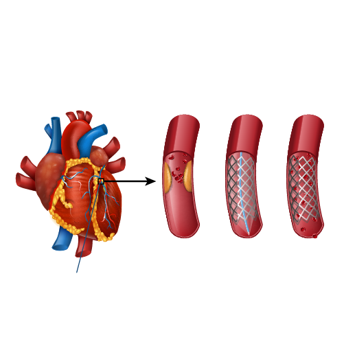
Angioplasty
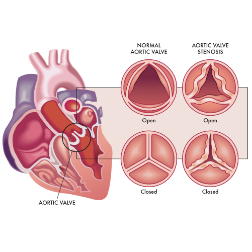
Aortic Stenosis
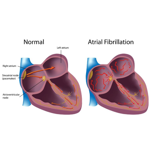
Atrial Fibrillation
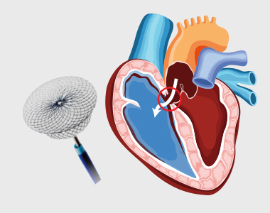
Atrial Septal Defect
Related Posts